


Understanding Neural Control and Coordination for NEET
The Neural Control and Coordination is a crucial concept for the NEET Exam, covering the central and peripheral nervous systems, neurons, nerve impulses, and reflexes. This Concept is not only fundamental to understanding biology but also key to scoring well in NEET. In this guide, we break down all the important concepts in simple, digestible segments, ideal for quick revision.
What is Neural Control and Coordination?
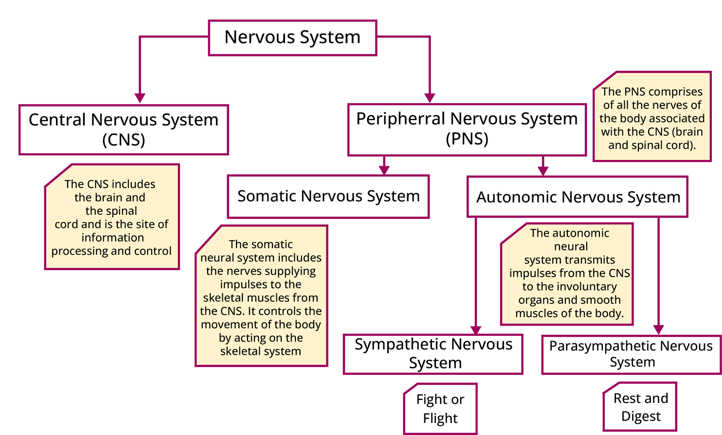
Neural Control and Coordination refer to the processes by which the nervous system and endocrine system work together to maintain the body’s functions. The nervous system is responsible for controlling and coordinating all voluntary and involuntary actions within the body, responding to external and internal stimuli. The central nervous system (CNS), consisting of the brain and spinal cord, and the peripheral nervous system (PNS), which connects the rest of the body to the CNS, are essential components in this process.
Nervous System
The neural system consists of specialised cells called neurons, which are capable of receiving and transmitting various signals. In simpler organisms like Hydra, a network of neurons is present. In insects, the neural system is made up of multiple ganglia and neural tissues. It is more advanced and complex in vertebrates.
Human Nervous System
The human nervous system consists of three main parts-
Central Nervous System (CNS) -In vertebrates, the CNS is hollow, dorsal, and lacks ganglia. In contrast, invertebrates have a ventral, solid CNS with ganglia. The CNS is divided into the brain and the spinal cord.
Peripheral Nervous System (PNS) -The PNS consists of nerves that extend between the CNS and body parts. It includes both cranial and spinal nerves, and it controls voluntary functions.
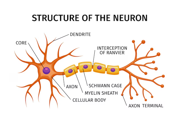
Neurons
Neurons are the fundamental units of the nervous system. They consist of three key parts-
Cell Body- Contains the cytoplasm, cell organelles, and Nissl’s granules.
Dendrites- Short fibers that branch out from the cell body and transmit impulses toward it.
Axon- A long fiber that transmits nerve impulses away from the cell body. The axon ends in synaptic knobs, which contain neurotransmitter-filled synaptic vesicles.
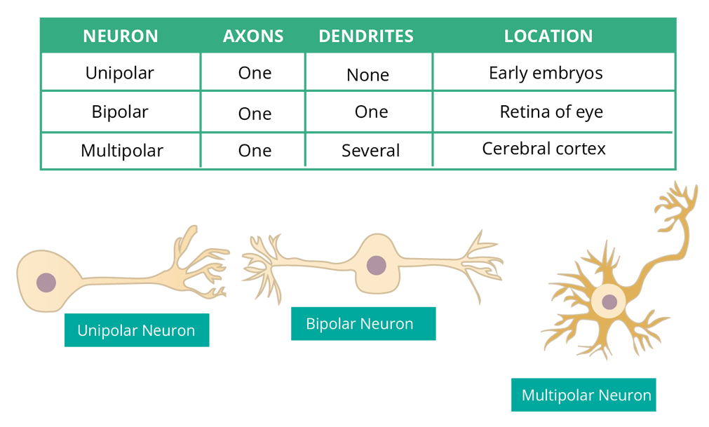
Types of Neurons
Based on the number of axons and dendrites, neurons are divided into three types, Unipolar, Bipolar, and Multipolar.
Nerve Impulse
Since the membranes of neurons are polarised, they are called excitable cells.
Membranes are polarised because there are different types of ion channels present on the neural membrane.
Different ions selectively pass through these ion channels.
The resting potential is defined as the electrical potential difference across the plasma membrane at rest.
The resting potential of the membrane is approx -70 mV.
When a neuron is not transmitting any impulse, it means it is in the resting phase. The membrane of the axon is more permeable to potassium ions (K+) and almost impermeable to sodium ions (Na+) when it is at rest.
Similarly, negatively charged proteins in the axoplasm are impermeable to the membrane.
As a result, the axoplasm within the axon has a high concentration of K+ and negatively charged proteins but a low quantity of Na+.
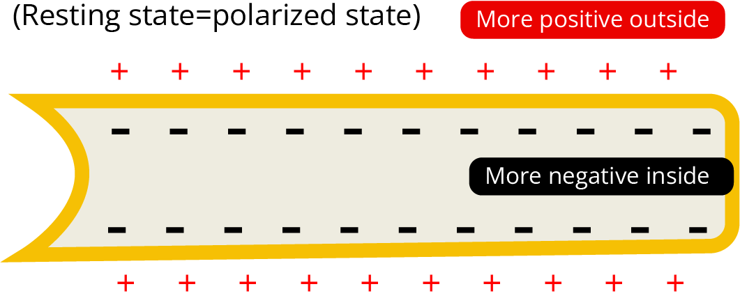
The fluid outside the axon, on the other hand, includes a low concentration of K+ and a high concentration of Na+, forming a concentration gradient.
The active transport of ions by the sodium-potassium pump, which transfers 3 Na+ outside for 2K+ into the cell, maintains these ionic gradients across the resting membrane.
As a result, the axonal membrane's outer surface has a positive charge, whereas its inner surface has a negative charge and is, therefore, polarised.
The resting potential is defined as the difference in electrical potential across the plasma membrane at rest.
Impulse Generation and Conduction
The stimulus delivered at point A on the polarised membrane becomes freely permeable to Na+.
This causes a fast influx of Na+, followed by polarity reversal at that site. (outside negative and inside positive-depolarisation, approx. +30mV)
At sites immediately ahead, the membrane has a positive charge on the outer surface and a negative charge on its inner surface.
Now, the current flows on the inner surface from site A to site B and at site B, the polarity is reversed, and an action potential is formed. As a result, the impulse (action potential) produced at site A reaches Site B.
The process is repeated along the length of the axon, and the impulse is transmitted as a result.
Transmission of Nerve Impulse
At the point where a chemical synapse forms, the membranes of the pre- and postsynaptic neurons are separated at the synaptic cleft.
Here, neurotransmitters (chemicals) are involved in impulse transmission.
The migration of synaptic vesicles towards the membrane is stimulated when an impulse (action potential) arrives at the axon terminal and synaptic vesicles fuse with the plasma membrane releasing neurotransmitters in the synaptic cleft.
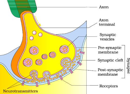
The influx of Ca2+ ions in the presynaptic cleft helps in the movement of synaptic vesicles.
The released neurotransmitters from synaptic vesicles bind to their specific receptors, present on the postsynaptic membrane.
This binding opens ion channels, allowing ions to enter the postsynaptic neuron and generate a new potential.
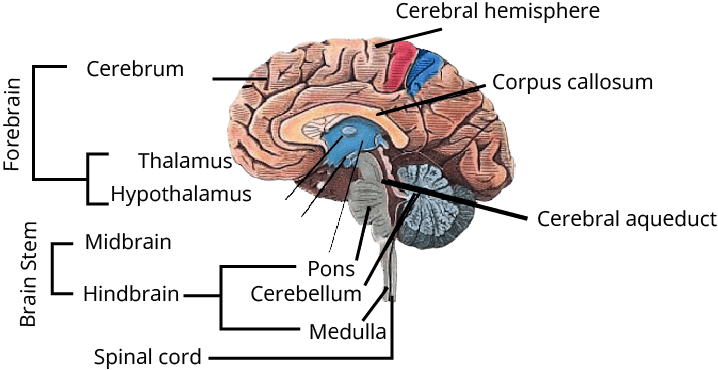
Human Brain
The forebrain, midbrain, and hindbrain are the three major parts of the brain.
The cerebrum, thalamus, and hypothalamus make up the forebrain.
The midbrain is situated between the forebrain's thalamus/hypothalamus and the pons of the hindbrain.
The pons, cerebellum, and medulla make up the hindbrain (also called the medulla oblongata).
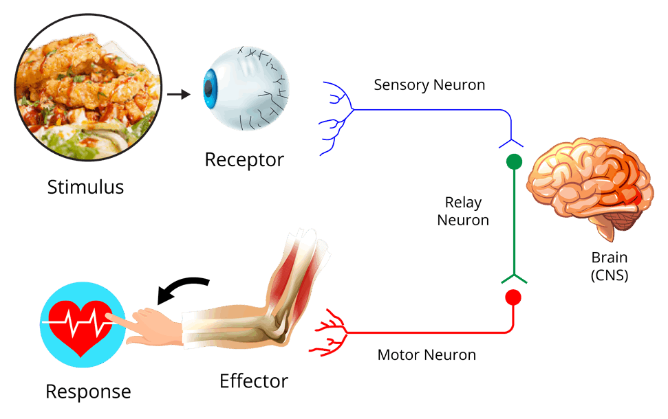
Reflex Action and Reflex Arc
Reflex Action is an involuntary response to a stimulus, mediated by a specific pathway in the Central Nervous System (CNS). This pathway includes both afferent neurons and efferent neurons-
Afferent NeuronsThese neurons receive signals from sensory organs and carry them to the CNS.
Efferent Neurons- These neurons transmit signals from the CNS to the effector organ, causing a response.
A reflex arc is the route taken by the nerve impulses during this process, enabling a quick and automatic reaction to stimuli without conscious thought.
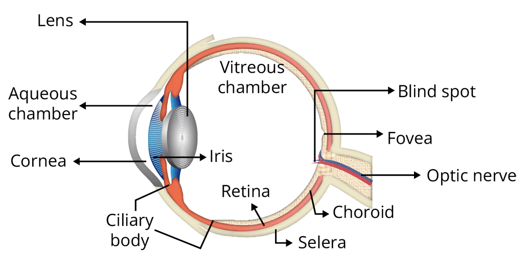
Human Eye
The human eye is a crucial organ for vision and consists of several key structures-
Eyeball Layers-
External Layer- Made of sclera, a dense connective tissue. The front part of the sclera is the cornea.
Middle Layer- Known as the choroid, which is rich in blood vessels. The ciliary body extends forward, leading to the iris, which is pigmented and opaque.
Lens- The eye contains a transparent lens held in place by ligaments attached to the ciliary body. The pupil, a small aperture in the center of the iris, controls the amount of light entering the eye by adjusting its diameter through the iris muscles.
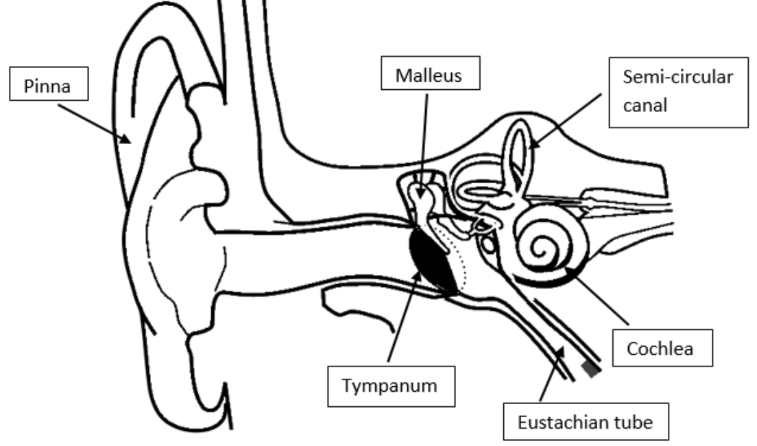
Human Ear
The human ear plays two main roles- hearing and balance regulation. It is divided into three sections-
Outer Ear-
Composed of the pinna and external auditory meatus.
The pinna captures sound vibrations, while the external auditory meatus directs them to the eardrum.
Middle Ear-
Contains three ossicles- malleus, incus, and stapes, which are connected.
The middle ear is linked to the pharynx via the eustachian tube, helping balance the pressure on both sides of the eardrum.
Inner Ear-
Known as the labyrinth, the inner ear consists of the bony labyrinth and the membranous labyrinth, essential for both hearing and balance.
Essential Study Materials for NEET UG Success
Neural Control and Coordination- Functions, Neurons, and Reflexes

 Share
ShareFAQs on Neural Control and Coordination- Functions, Neurons, and Reflexes
1. What is Neural Control and Coordination?
Neural control and coordination refer to the process by which the nervous system and endocrine system work together to regulate and control the functions of the body. It involves the transmission of electrical signals through neurons, which helps in coordinating voluntary and involuntary actions.
2. How does the nervous system coordinate voluntary actions?
The nervous system coordinates voluntary actions through the somatic nervous system. This system transmits signals from the central nervous system (CNS) to the skeletal muscles, enabling conscious control over movements such as walking, writing, or speaking.
3. What is the role of neurons in neural coordination?
Neurons are the building blocks of neural control and coordination. They transmit electrical impulses across the body, helping to communicate signals between different parts of the body and the brain. There are three main parts of a neuron- the cell body, dendrites, and axon- each playing a crucial role in transmitting and processing these signals.
4. What is a reflex action in neural control?
A reflex action is an involuntary and automatic response to a stimulus. It is a quick and immediate reaction that does not require conscious thought. Reflex actions are controlled by the spinal cord and are part of the neural control system.
5. What is the difference between the central nervous system and the peripheral nervous system?
The central nervous system (CNS) consists of the brain and spinal cord, which process and interpret sensory information. The PNS connects the CNS to the rest of the body through nerves. The PNS includes cranial nerves and spinal nerves and controls the voluntary and involuntary actions of the body.
6. How do neurotransmitters play a role in neural coordination?
Neurotransmitters are chemicals that transmit signals across synapses between neurons. They play a crucial role in neural coordination, ensuring that electrical impulses are passed from one neuron to the next, which is vital for the functioning of the nervous system and the coordination of bodily activities.
7. What is the function of the myelin sheath in neural control?
The myelin sheath is a fatty layer that wraps around the axons of some neurons. It acts as an insulator, speeding up the transmission of electrical signals. This facilitates faster and more efficient neural coordination in the body.
8. What are reflex arcs in the nervous system?
A reflex arc is the neural pathway that controls reflex actions. It typically involves a sensory neuron, an interneuron (in the spinal cord), and a motor neuron. Reflex arcs allow for immediate, involuntary responses to stimuli, bypassing the brain for quicker reactions.
9. How does the autonomic nervous system contribute to neural control?
The autonomic nervous system (ANS) is responsible for controlling involuntary functions, such as heart rate, digestion, and respiratory rate. It operates automatically without conscious control and plays a vital role in maintaining homeostasis in the body through neural control.
10. Can the nervous system repair itself after injury?
The nervous system has a limited capacity for repair. Peripheral nerves (outside the brain and spinal cord) can regenerate to some extent, while the central nervous system has very little regenerative ability. However, research in neural regeneration continues to explore ways to repair and regenerate damaged neural tissues.
11. What is the basic unit of the nervous system?
The neuron is the fundamental unit of the nervous system. Neurons are specialised cells responsible for transmitting electrical and chemical signals throughout the body, enabling various functions such as sensation, movement, and thought.
12. How does an impulse travel across a synapse?
When an electrical impulse reaches the end of a neuron (axon terminal), it triggers the release of neurotransmitters into the synaptic cleft (the gap between two neurons). These neurotransmitters bind to receptors on the post-synaptic neuron, initiating a new electrical impulse in that neuron.
13. What is saltatory conduction?
Saltatory conduction refers to the process by which nerve impulses jump from one node of Ranvier to the next along a myelinated axon. This mechanism significantly speeds up the transmission of electrical signals in the nervous system.
14. How many pairs of cranial nerves are present in humans?
Humans have 12 pairs of cranial nerves. These nerves emerge directly from the brain and are primarily responsible for the sensory and motor functions of the head and neck.