


Draw a labelled diagram of the T.S. of the spinal cord of man.
Answer
561.6k+ views
Hint: The spinal cord is a thin, long, and tube-like structure composed of nervous tissue that starts from the medulla oblongata and terminates at the lumbar region of the vertebral column, and circles the central canal that has the cerebrospinal fluid (CSF). The brain and the spinal cord form the central nervous system (CNS) of the body.
Complete answer:
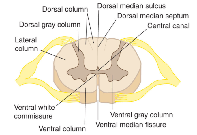
The spinal cord in humans starts with the occipital bone, passes through the foramen magnum, and then goes inside the spinal canal at the cervical vertebrae. The spinal cord is covered with three layers known as meninges. The outermost layer is called the dura mater, the middle one is the arachnoid mater, and the innermost is called the pia mater. The cerebrospinal fluid is present between the arachnoid and pia mater. Just like the brain, the spinal cord is also made up of white and gray matter. The gray matter looks like a butterfly and is present at the center of the spinal cord (as shown in the diagram). It is composed of neuronal cell bodies. It has dorsal and ventral horns. The grey matter is surrounded by white matter which is composed of axons. It contains the paths to connect the brain with the other parts of the body. Blood supply is through the vertebral artery that gives rise to the anterior and posterior spinal arteries. On the dorsal root of the spinal cord, the spinal ganglion is located.
Note: The anatomy of the spinal cord is according to the function it performs. The major function of the spinal cord is to conduct the impulses from the brain to the body and thereby, producing reflexes. It transmits the signal from the motor cortex to the other parts of the body and from afferent fibers to the sensory cortex.
Complete answer:
The spinal cord in humans starts with the occipital bone, passes through the foramen magnum, and then goes inside the spinal canal at the cervical vertebrae. The spinal cord is covered with three layers known as meninges. The outermost layer is called the dura mater, the middle one is the arachnoid mater, and the innermost is called the pia mater. The cerebrospinal fluid is present between the arachnoid and pia mater. Just like the brain, the spinal cord is also made up of white and gray matter. The gray matter looks like a butterfly and is present at the center of the spinal cord (as shown in the diagram). It is composed of neuronal cell bodies. It has dorsal and ventral horns. The grey matter is surrounded by white matter which is composed of axons. It contains the paths to connect the brain with the other parts of the body. Blood supply is through the vertebral artery that gives rise to the anterior and posterior spinal arteries. On the dorsal root of the spinal cord, the spinal ganglion is located.
Note: The anatomy of the spinal cord is according to the function it performs. The major function of the spinal cord is to conduct the impulses from the brain to the body and thereby, producing reflexes. It transmits the signal from the motor cortex to the other parts of the body and from afferent fibers to the sensory cortex.
Recently Updated Pages
Master Class 11 Computer Science: Engaging Questions & Answers for Success
Master Class 11 Business Studies: Engaging Questions & Answers for Success
Master Class 11 Economics: Engaging Questions & Answers for Success
Master Class 11 English: Engaging Questions & Answers for Success
Master Class 11 Maths: Engaging Questions & Answers for Success
Master Class 11 Biology: Engaging Questions & Answers for Success
Trending doubts
One Metric ton is equal to kg A 10000 B 1000 C 100 class 11 physics CBSE
There are 720 permutations of the digits 1 2 3 4 5 class 11 maths CBSE
Discuss the various forms of bacteria class 11 biology CBSE
Draw a diagram of a plant cell and label at least eight class 11 biology CBSE
State the laws of reflection of light
10 examples of friction in our daily life