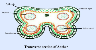
Draw the diagram of microsporangium of an Angiosperm and label any four parts. State the function of its innermost wall layer.
Answer
540.6k+ views
Hint: Microsporangium is a sac-like structure consisting of microspores which form pollen grains. Microsporangium is divided into the microsporangium wall and sporogenous tissue in which microsporangium wall is divided into the epidermis, endothecium, middle layer and tapetum.
Complete answer:

Microsporangium of an Angiosperm
The anther of an angiosperm is generally bilobed in a structure which consists of microsporangium. This microsporangium consists of microsporangia which divide to form pollen grains. The microsporangium consists of four wall layers outside and sporogenous tissue inside. The microsporangium wall is divided into outer layer epidermis, hypodermal wall layer endothecium, middle layer and innermost nourishing layer tapetum. Tapetum is the innermost layer consisting of a dense cytoplasm which after endomitosis becomes enlarged in size and multinucleate. The tapetum cell performs various functions like it provides nourishment to the developing sporogenous tissue, microspore mother cell and microspore. It helps in the binding of microspore, by the production of enzymes callose. It secretes ubisch granules which provide a covering to the pollen grains. It provides a covering to entomophilous pollen grain with the help of pollenkitt, which is adhesive, provides odour which helps to attract insects. It also provides protein which helps to find out compatible and incompatible pollen grains. It also releases enzymes, hormones, amino acids, etc during microsporogenesis which help in the development of young pollen.
Note:
The ubisch bodies of pollen grains are also known as orbicules. It occurs in different shapes and is made up of lipids. It does not perform specific functions but acts as a by-product of tapetal cell metabolism. In some cases, ubisch bodies help in the lysis of tapetal cells.
Complete answer:

Microsporangium of an Angiosperm
The anther of an angiosperm is generally bilobed in a structure which consists of microsporangium. This microsporangium consists of microsporangia which divide to form pollen grains. The microsporangium consists of four wall layers outside and sporogenous tissue inside. The microsporangium wall is divided into outer layer epidermis, hypodermal wall layer endothecium, middle layer and innermost nourishing layer tapetum. Tapetum is the innermost layer consisting of a dense cytoplasm which after endomitosis becomes enlarged in size and multinucleate. The tapetum cell performs various functions like it provides nourishment to the developing sporogenous tissue, microspore mother cell and microspore. It helps in the binding of microspore, by the production of enzymes callose. It secretes ubisch granules which provide a covering to the pollen grains. It provides a covering to entomophilous pollen grain with the help of pollenkitt, which is adhesive, provides odour which helps to attract insects. It also provides protein which helps to find out compatible and incompatible pollen grains. It also releases enzymes, hormones, amino acids, etc during microsporogenesis which help in the development of young pollen.
Note:
The ubisch bodies of pollen grains are also known as orbicules. It occurs in different shapes and is made up of lipids. It does not perform specific functions but acts as a by-product of tapetal cell metabolism. In some cases, ubisch bodies help in the lysis of tapetal cells.
Recently Updated Pages
Master Class 11 Computer Science: Engaging Questions & Answers for Success

Master Class 11 Business Studies: Engaging Questions & Answers for Success

Master Class 11 Economics: Engaging Questions & Answers for Success

Master Class 11 English: Engaging Questions & Answers for Success

Master Class 11 Maths: Engaging Questions & Answers for Success

Master Class 11 Biology: Engaging Questions & Answers for Success

Trending doubts
One Metric ton is equal to kg A 10000 B 1000 C 100 class 11 physics CBSE

There are 720 permutations of the digits 1 2 3 4 5 class 11 maths CBSE

Discuss the various forms of bacteria class 11 biology CBSE

Draw a diagram of a plant cell and label at least eight class 11 biology CBSE

State the laws of reflection of light

Explain zero factorial class 11 maths CBSE


