


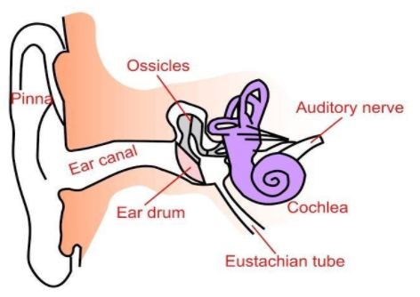
Draw the diagram showing the internal structure of the human ear and label the following parts.
A. Organ of Corti
B. Auditory nerve
Answer
585.9k+ views
Hint: Ears are the sensory organs that detect and receive the sound waves and convert them into the nerve impulses, hence help in hearing. However hearing is not the sole function of the ears. The ears also maintain the body balance or body equilibrium. So, the ears perform two sensory functions: Hearing, and Maintenance of body balance
Complete answer:
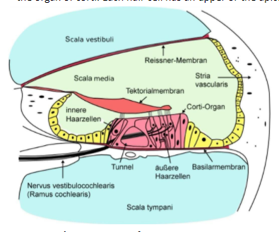
Organ of corti:
The Organ of Corti is a structure located on the basilar membrane, first described by Italian microscopist, Alfonso Corti (1822-1888). It is a sensory structure which contains hair cells. The hair cells act as the auditory receptors. The hair cells are present in rows on the internal side of the organ of corti. Each hair cell has an upper or the apical end and lower or the basal end.
Auditory nerve :
The outer ear consists of the pinna and external auditory meatus that leads to a canal. Comes the external auditory meatus and canal. (Auditory term comes in the relation of the sound). The canal carries the sound waves, that is why it is called the external auditory canal. The sound waves, present in the air, are collected by the pinna and carried inwards by the external auditory canal.
Note: The hair cells are present in rows on the internal side of the organ of corti. Each hair cell has an upper or the apical end and lower or the basal end. A large number of processes (projected parts) called stereocilia. Tectorial membrane is a delicate and flexible membrane that covers the tips or apices of the hair cells
Complete answer:
Organ of corti:
The Organ of Corti is a structure located on the basilar membrane, first described by Italian microscopist, Alfonso Corti (1822-1888). It is a sensory structure which contains hair cells. The hair cells act as the auditory receptors. The hair cells are present in rows on the internal side of the organ of corti. Each hair cell has an upper or the apical end and lower or the basal end.
Auditory nerve :
The outer ear consists of the pinna and external auditory meatus that leads to a canal. Comes the external auditory meatus and canal. (Auditory term comes in the relation of the sound). The canal carries the sound waves, that is why it is called the external auditory canal. The sound waves, present in the air, are collected by the pinna and carried inwards by the external auditory canal.
Note: The hair cells are present in rows on the internal side of the organ of corti. Each hair cell has an upper or the apical end and lower or the basal end. A large number of processes (projected parts) called stereocilia. Tectorial membrane is a delicate and flexible membrane that covers the tips or apices of the hair cells
Recently Updated Pages
Master Class 11 Computer Science: Engaging Questions & Answers for Success
Master Class 11 Business Studies: Engaging Questions & Answers for Success
Master Class 11 Economics: Engaging Questions & Answers for Success
Master Class 11 English: Engaging Questions & Answers for Success
Master Class 11 Maths: Engaging Questions & Answers for Success
Master Class 11 Biology: Engaging Questions & Answers for Success
Trending doubts
One Metric ton is equal to kg A 10000 B 1000 C 100 class 11 physics CBSE
There are 720 permutations of the digits 1 2 3 4 5 class 11 maths CBSE
Discuss the various forms of bacteria class 11 biology CBSE
Draw a diagram of a plant cell and label at least eight class 11 biology CBSE
State the laws of reflection of light
Explain zero factorial class 11 maths CBSE