In the gastrointestinal tract, the Meissner’s plexus and Auerbach’s plexus occur respectively in the,
(a) Lamina Propria and muscularis mucosa
(b) Submucosa and muscularis externa
(c) Submucosa and mucosa
(d) Mucosa and muscularis externa
Answer
571.8k+ views
Hint: Plexus is the branching and networking of vessels or nerves in a particular region. The Meissner’s plexus and Auerbach’s plexus are present in the second, and third layers of the stomach wall where the contents are fibrous connective tissues and smooth muscles respectively.
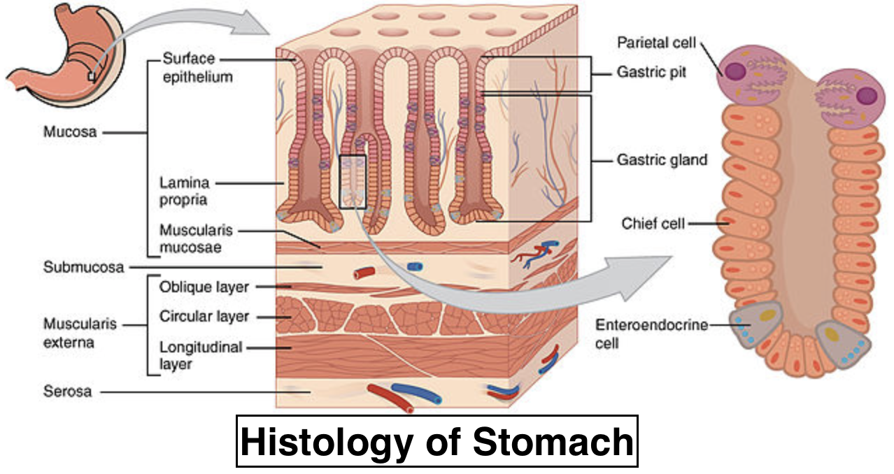
Complete answer: In the gastrointestinal tract, the Meissner’s plexus and Auerbach’s plexus occur in the Submucosa and muscularis externa respectively. There are four layers of stomach walls namely, Mucosa, Submucosa, Muscularis externa, and Serosa (from deep to superficial).
Muscularis Externa: The smooth muscle situated deeply in the submucosa is the gastric muscularis externa. It is composed of 3 layers: oblique inside, circular center, and longitudinal outside. Churning movements necessary for mechanical digestion are generated by the muscularis externa layer. They cast the mucosa and submucosa into rugae as these layers contract. The myenteric (Auerbach's) plexus is located within the muscularis externa, bearing both sympathetic and parasympathetic fibers to the smooth muscle layers. Via interstitial cells from Cajal (ICCs), the neurons of this plexus are connected to smooth muscle cells. Such cells act as intrinsic pacemakers of the gut, regulating the slow contractions of the stomach wall necessary for food churning, as well as mediating neural signals. The operation of ICCs is regulated by the autonomic nervous system.

The submucosa layer of the stomach is situated around the mucosa. Various connective tissues, blood vessels, and nerves are made up of the submucosa. Connective tissues support the mucosal tissues and attach them to the layer of muscularis. The submucosa's blood supply supplies the wall of the stomach with nutrients. The contents of the stomach are monitored by nervous tissue in the submucosa and smooth muscle contraction and digestive substance secretion are managed. It contains the Meissner’s plexus that carries parasympathetic fibers.
So, the answer is “Submucosa and muscularis externa”.
Additional Information: The stomach, sitting between the esophagus and duodenum, is a vital part of the gastrointestinal (GI) tract. Its functions are to combine food with stomach acid and, utilizing chemical and mechanical digestion, break food down into smaller particles.
The outer layer is smooth, continuous with the parietal peritoneum of the stomach wall. The inner wall (layers of mucosa and submucosa) is thrown into folds known as rugae, or gastric folds, causing the stomach to distend after the food enters.
The gastric mucosa is the innermost layer of the stomach wall. It is created by a surface epithelium layer and a lamina propria and muscularis mucosae underlying it.
The outermost layer of the stomach wall is Gastric serosa. It consists of a layer, known as mesothelium, of simple squamous epithelium, and a thin layer of connective tissue underlying it.
Note: - Auerbach’s plexus is also known as mesenteric plexus. - In all regions of the intestine and the entire gastrointestinal tract, this layered arrangement follows the same general structure. - The Gastric muscularis externa is also known as Tunica muscularis. - In the stomach, there are three types of glands; cardiac, gastric, and pyloric, named for the area in which they are located. - Mnemonic for the four layers of stomach is M.S.M.S (Mucosa, Muscularis externa, Submucosa, and Serosa) - Mnemonic for the plexus: S.M.P (Submucosal, Meissner, Parasympathetic) M.A.P.S (Muscularis externa, Auerbach’s, Parasympathetic, Sympathetic)
Recently Updated Pages
Master Class 11 Computer Science: Engaging Questions & Answers for Success

Master Class 11 Business Studies: Engaging Questions & Answers for Success

Master Class 11 Economics: Engaging Questions & Answers for Success

Master Class 11 English: Engaging Questions & Answers for Success

Master Class 11 Maths: Engaging Questions & Answers for Success

Master Class 11 Biology: Engaging Questions & Answers for Success

Trending doubts
One Metric ton is equal to kg A 10000 B 1000 C 100 class 11 physics CBSE

There are 720 permutations of the digits 1 2 3 4 5 class 11 maths CBSE

Discuss the various forms of bacteria class 11 biology CBSE

Draw a diagram of a plant cell and label at least eight class 11 biology CBSE

State the laws of reflection of light

Explain zero factorial class 11 maths CBSE


