


In the given diagram of a section of bone tissue, certain parts have been indicated by alphabets. Select the answer in which these alphabets have been correctly matched with the parts which they include.
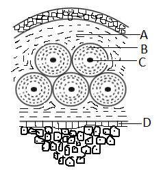
A) A- Interstitial lamellae; B- Nerve; C- Canaliculi; D- Haversian Canal
B) A- Interstitial lamellae; B- Osteocytes; C- Harvesian system; D- Canaliculi
C) A- Interstitial lamellae; B- Osteocytes; C- Nerve; D- Blood vessels
D) A- Interstitial lamellae; B- Osteocytes; C- Harvesian canal; D- Endosteum
Answer
561.6k+ views
Hint: The skeletal system is formed of bones and bones themselves are formed of various elements. These are bone tissue, periosteum, yellow and red bone marrow, and endosteum. There are two bone tissues named compact bone tissue and spongy bone tissue. The given figure is of compact bone tissue. It forms the outer layer of bones.
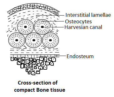
Complete answer: The compact bone tissue forms the outermost layer. The tissue provides protection and support to the bone. It also helps the bones to bear stress and weight put on them by the body. Heavy physical work puts pressure on these tissues as they maintain the weight. The very basic unit of compact bone tissue is osteon. It is also known as the Haversian system. Each of the osteon is made up of a cylindrical structure made of four parts. The central tube that contains blood vessels and nerves is called the Haversian canal. It is represented by ‘C’ in the figure. The concentric ring composed of a strong matrix is called the interstitial lamellae. It is shown by A in the figure. It is formed from mineral salts like calcium and phosphate and collagen fibres. The mineral salts provide hardness to the bone structure. The collagen fibres help to provide strength to the bone. The small space present between the interstitial lamellae is called the lacunae. It contains bone cells called osteocytes. Osteocytes are represented by ‘B’ in the figure. The ‘D’ in the figure represents Endosteum. It is a thin vascular lining of vascular tissue. It is present as the inner lining of the bony tissue. It forms the medullary cavity of long bones.
So, the right answer is A-Interstitial lamellae; B-Osteocytes; C-Harvesian canal; D-Endosteum.
Thus, option D is the correct answer.
Note: The bone tissues provide support to softer tissues of the skeleton. It provides mechanical protection reducing the risks of internal injury. For example, vertebrae protect the spinal cord from injury, the cranium protects the brain from injury, etc.
Recently Updated Pages
Master Class 11 Computer Science: Engaging Questions & Answers for Success
Master Class 11 Business Studies: Engaging Questions & Answers for Success
Master Class 11 Economics: Engaging Questions & Answers for Success
Master Class 11 English: Engaging Questions & Answers for Success
Master Class 11 Maths: Engaging Questions & Answers for Success
Master Class 11 Biology: Engaging Questions & Answers for Success
Trending doubts
One Metric ton is equal to kg A 10000 B 1000 C 100 class 11 physics CBSE
There are 720 permutations of the digits 1 2 3 4 5 class 11 maths CBSE
Discuss the various forms of bacteria class 11 biology CBSE
Draw a diagram of a plant cell and label at least eight class 11 biology CBSE
State the laws of reflection of light
Explain zero factorial class 11 maths CBSE