


Nissl's granules are found in the cyton of nerve cells. These have an affinity for basic dyes. The granules are made up of
A. Proteins
B. DNA
C. Amino acids
D. Rough Endoplasmic Reticulum
Answer
603.3k+ views
Hint: These particles are present in the cell body of the neurons and responsible for the synthesis of proteins.
Complete answer:
A Nissl body is a large granular body which is made up of rough endoplasmic reticulum. It consists of rosettes of free ribosomes, and are the site of protein synthesis. The proteins flow along the dendrites and the axon and replace the proteins which are broken down during cellular activities.
Additional information:
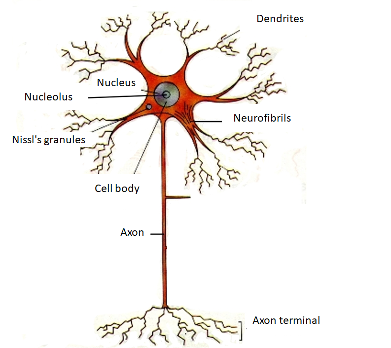
- A neuron is a specialized cell that transmits nerve impulses from one nerve cell to another via synapses.
- Numerous microscopic clumps called Nissl bodies are seen when nerve cells are stained with basophilic dye.
- Basophilic dye highlights negatively charged components and binds to the phosphate backbone of the ribosomal RNA.
- Nissl granule was named after a German neuropathologist Franz Nissl, who invented the Nissl staining method.
- The functions of Nissl bodies are thought to be the same as that of the rest of the endoplasmic reticulum and the Golgi apparatus, which is the release and manufacture of proteins and amino acids.
So, the correct answer is ‘Rough Endoplasmic Reticulum’.
Note: A typical neuron consists of a cell body (soma), dendrites, and a single axon. The axon and the dendrites are the filamentous structure while soma is the compact cell body. The axons send the signals downwards and out of the nerve cells which are received by the dendrites along with the soma. The Nissl granules are found in the soma of the neuron.
Complete answer:
A Nissl body is a large granular body which is made up of rough endoplasmic reticulum. It consists of rosettes of free ribosomes, and are the site of protein synthesis. The proteins flow along the dendrites and the axon and replace the proteins which are broken down during cellular activities.
Additional information:
- A neuron is a specialized cell that transmits nerve impulses from one nerve cell to another via synapses.
- Numerous microscopic clumps called Nissl bodies are seen when nerve cells are stained with basophilic dye.
- Basophilic dye highlights negatively charged components and binds to the phosphate backbone of the ribosomal RNA.
- Nissl granule was named after a German neuropathologist Franz Nissl, who invented the Nissl staining method.
- The functions of Nissl bodies are thought to be the same as that of the rest of the endoplasmic reticulum and the Golgi apparatus, which is the release and manufacture of proteins and amino acids.
So, the correct answer is ‘Rough Endoplasmic Reticulum’.
Note: A typical neuron consists of a cell body (soma), dendrites, and a single axon. The axon and the dendrites are the filamentous structure while soma is the compact cell body. The axons send the signals downwards and out of the nerve cells which are received by the dendrites along with the soma. The Nissl granules are found in the soma of the neuron.
Recently Updated Pages
Master Class 11 Computer Science: Engaging Questions & Answers for Success
Master Class 11 Business Studies: Engaging Questions & Answers for Success
Master Class 11 Economics: Engaging Questions & Answers for Success
Master Class 11 English: Engaging Questions & Answers for Success
Master Class 11 Maths: Engaging Questions & Answers for Success
Master Class 11 Biology: Engaging Questions & Answers for Success
Trending doubts
One Metric ton is equal to kg A 10000 B 1000 C 100 class 11 physics CBSE
There are 720 permutations of the digits 1 2 3 4 5 class 11 maths CBSE
Discuss the various forms of bacteria class 11 biology CBSE
Draw a diagram of a plant cell and label at least eight class 11 biology CBSE
State the laws of reflection of light
10 examples of friction in our daily life