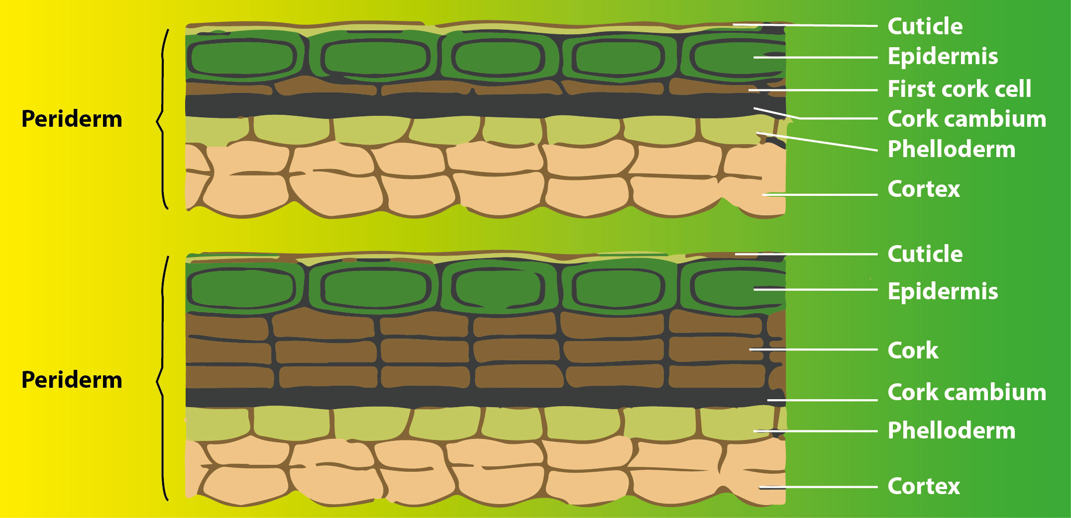
Periderm is formed from
(a) Vascular cambium
(b) Phellogen
(c) Fascicular cambium
(d) Interfascicular cambium
Answer
565.5k+ views
Hint: It refers to the collective term wanting to signify phellogen, phellem, and phelloderm during a plant stem. It's formed in plants, mainly occurring to exchange the prevailing epidermis. It's composed of thin-walled, narrow, and rectangular cells.
Complete answer:
A. Vascular cambium – produces secondary vascular tissues and ray parenchyma.
B. Fascicular cambium – a strip of cambium present between the xylem and phloem of a vascular strand is named fascicular cambium. It involves secondary growth.
C. Phellogen – a hoop of cambium formed within the cortex of dicot stem, by the dedifferentiation of parenchyma is named Phellogen. it's involved within the formation of the periderm.
D. Intrafascicular cambium - a strip of cambium present between the xylem and phloem of a vascular strand is named fascicular cambium. It involves secondary growth.
Additional information: The formation of periderm occurs during secondary growth. During this process, to exchange the broken outer epidermal layer and therefore the cortical layer, the cells of the cortex turn meristematic.

Thus, cork cambium or phellogen is made. The phellogen comprises slightly walled, restricted, and rectangular cells. Later on, the phellogen cuts off cells on either side. The cells of the external side produce the phellem or plug which because of the statement of suberin in its cell layer is impenetrable to water. Additionally, the inward side structures the optional cortex or phelloderm which is particularly parenchymatous in nature.
So the correct answer is ‘Phellogen’.
Note: Analysis of mature tuber periderm, however, might not produce easily identifiable phellogen or phelloderm. A study of all three periderm cell types in immature and mature periderm was needed to work out maturational changes. Recently have these periderm cell structures been plainly shown for simpler identification and related morphological depiction. The suberization measures engaged with phellem improvement are just halfway described.
Complete answer:
A. Vascular cambium – produces secondary vascular tissues and ray parenchyma.
B. Fascicular cambium – a strip of cambium present between the xylem and phloem of a vascular strand is named fascicular cambium. It involves secondary growth.
C. Phellogen – a hoop of cambium formed within the cortex of dicot stem, by the dedifferentiation of parenchyma is named Phellogen. it's involved within the formation of the periderm.
D. Intrafascicular cambium - a strip of cambium present between the xylem and phloem of a vascular strand is named fascicular cambium. It involves secondary growth.
Additional information: The formation of periderm occurs during secondary growth. During this process, to exchange the broken outer epidermal layer and therefore the cortical layer, the cells of the cortex turn meristematic.

Thus, cork cambium or phellogen is made. The phellogen comprises slightly walled, restricted, and rectangular cells. Later on, the phellogen cuts off cells on either side. The cells of the external side produce the phellem or plug which because of the statement of suberin in its cell layer is impenetrable to water. Additionally, the inward side structures the optional cortex or phelloderm which is particularly parenchymatous in nature.
So the correct answer is ‘Phellogen’.
Note: Analysis of mature tuber periderm, however, might not produce easily identifiable phellogen or phelloderm. A study of all three periderm cell types in immature and mature periderm was needed to work out maturational changes. Recently have these periderm cell structures been plainly shown for simpler identification and related morphological depiction. The suberization measures engaged with phellem improvement are just halfway described.
Recently Updated Pages
Master Class 11 Computer Science: Engaging Questions & Answers for Success

Master Class 11 Business Studies: Engaging Questions & Answers for Success

Master Class 11 Economics: Engaging Questions & Answers for Success

Master Class 11 English: Engaging Questions & Answers for Success

Master Class 11 Maths: Engaging Questions & Answers for Success

Master Class 11 Biology: Engaging Questions & Answers for Success

Trending doubts
One Metric ton is equal to kg A 10000 B 1000 C 100 class 11 physics CBSE

There are 720 permutations of the digits 1 2 3 4 5 class 11 maths CBSE

Discuss the various forms of bacteria class 11 biology CBSE

Draw a diagram of a plant cell and label at least eight class 11 biology CBSE

State the laws of reflection of light

Explain zero factorial class 11 maths CBSE


