


What is the structure and function of the compact bone?
Answer
485.7k+ views
Hint: Bone is not completely a solid structure but has many small hollow spaces between its cells and extracellular matrix components. Some of the spaces serve as blood vessel channels that supply nutrition to the bone tissue. Other areas serve for the space accommodation of the red and yellow bone marrows.
Depending on the size and distribution of the spaces the regions of a bone may be categorized as compact or spongy. Overall, about 80% of the skeleton comprises the compact bone and the rest 20% comprises the spongy bone.
Complete answer:
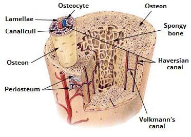
The structural aspects of compact bone tissue are:
1. It contains a few spaces and is the strongest form of bone tissue. It is found beneath the periosteum of all bones and makes up the bulk of the diaphysis of long bones.
2. The compact bone tissues are made up of repeating structural units called osteons, or haversian systems.
3. Each osteon consists of concentric lamellae arranged around a central canal or haversian canal. Between the concentric lamellae there are small spaces located that contain osteocytes. The neighboring osteocytes communicate via gap junctions.
4. These tube-like units of bone generally form a series of parallel cylinders that tend to run parallel to the axis of bones.
5. Small channels called canaliculi radiate along all directions from the lamellae. These tiny channels contain extracellular fluid. These also contain osteocytes.
6. The canaliculi connect the different lacunae with each other and together with the central canal form an intricate, miniature system of interconnected canals throughout the bone. This system serves the purpose of providing nutrients and oxygen to the bony tissue and removal of wastes.
7. The areas between the neighboring osteons contain lamellae called interstitial lamellae. These are actually fragments of older osteons that have been partially destroyed during bone rebuilding, growth and in case of deformations like fractures.
8. The blood vessels and nerves enter the compact bone through transverse perforating canals or Volkmann’s canals. The vessels and the nerves of these canals connect with those of the medullary cavity, periosteum, and central canals.
9. Arranged around the entire outer and inner circumference of the shaft of a long bone is the circumferential lamellae. They develop during initial bone formation and are connected to the periosteum via perforating fibers or Sharpey’s fibers.
Below is a diagram showing a detailed view of the internal structure of a compact bone:
Now, the functions of the compact bone are:
1. The compact bone tissue provides protection and support and resist the stresses produced by weight and movement. It is the major bone component of the long bones of the hand and legs which support most of the body weight.
2. It serves as storage areas for the red bone marrow.
Note:
The compact bone is actually present in those bones that are involved in stressful activities, like the bones of the hand and legs, ribs, etc. Osteons in compact bone are aligned generally in the same direction and are parallel to the length of the diaphysis. As a result, the shaft of a long bone resists bending or fracturing even when considerable force is applied from either end. Compact bones are thicker in those areas where the stress falls from many directions, not just in a particular line.
The lines of stress falling upon a bone are not static. They change as a person learns to walk and in response to repeated strenuous activities, and also due to fractures and physical deformities due to age. Thus, the organization of osteons is not static but changes overtime in response to the physical demands placed on the skeleton.
Depending on the size and distribution of the spaces the regions of a bone may be categorized as compact or spongy. Overall, about 80% of the skeleton comprises the compact bone and the rest 20% comprises the spongy bone.
Complete answer:
The structural aspects of compact bone tissue are:
1. It contains a few spaces and is the strongest form of bone tissue. It is found beneath the periosteum of all bones and makes up the bulk of the diaphysis of long bones.
2. The compact bone tissues are made up of repeating structural units called osteons, or haversian systems.
3. Each osteon consists of concentric lamellae arranged around a central canal or haversian canal. Between the concentric lamellae there are small spaces located that contain osteocytes. The neighboring osteocytes communicate via gap junctions.
4. These tube-like units of bone generally form a series of parallel cylinders that tend to run parallel to the axis of bones.
5. Small channels called canaliculi radiate along all directions from the lamellae. These tiny channels contain extracellular fluid. These also contain osteocytes.
6. The canaliculi connect the different lacunae with each other and together with the central canal form an intricate, miniature system of interconnected canals throughout the bone. This system serves the purpose of providing nutrients and oxygen to the bony tissue and removal of wastes.
7. The areas between the neighboring osteons contain lamellae called interstitial lamellae. These are actually fragments of older osteons that have been partially destroyed during bone rebuilding, growth and in case of deformations like fractures.
8. The blood vessels and nerves enter the compact bone through transverse perforating canals or Volkmann’s canals. The vessels and the nerves of these canals connect with those of the medullary cavity, periosteum, and central canals.
9. Arranged around the entire outer and inner circumference of the shaft of a long bone is the circumferential lamellae. They develop during initial bone formation and are connected to the periosteum via perforating fibers or Sharpey’s fibers.
Below is a diagram showing a detailed view of the internal structure of a compact bone:
Now, the functions of the compact bone are:
1. The compact bone tissue provides protection and support and resist the stresses produced by weight and movement. It is the major bone component of the long bones of the hand and legs which support most of the body weight.
2. It serves as storage areas for the red bone marrow.
Note:
The compact bone is actually present in those bones that are involved in stressful activities, like the bones of the hand and legs, ribs, etc. Osteons in compact bone are aligned generally in the same direction and are parallel to the length of the diaphysis. As a result, the shaft of a long bone resists bending or fracturing even when considerable force is applied from either end. Compact bones are thicker in those areas where the stress falls from many directions, not just in a particular line.
The lines of stress falling upon a bone are not static. They change as a person learns to walk and in response to repeated strenuous activities, and also due to fractures and physical deformities due to age. Thus, the organization of osteons is not static but changes overtime in response to the physical demands placed on the skeleton.
Recently Updated Pages
Master Class 11 Computer Science: Engaging Questions & Answers for Success
Master Class 11 Business Studies: Engaging Questions & Answers for Success
Master Class 11 Economics: Engaging Questions & Answers for Success
Master Class 11 English: Engaging Questions & Answers for Success
Master Class 11 Maths: Engaging Questions & Answers for Success
Master Class 11 Biology: Engaging Questions & Answers for Success
Trending doubts
One Metric ton is equal to kg A 10000 B 1000 C 100 class 11 physics CBSE
There are 720 permutations of the digits 1 2 3 4 5 class 11 maths CBSE
Discuss the various forms of bacteria class 11 biology CBSE
Draw a diagram of a plant cell and label at least eight class 11 biology CBSE
State the laws of reflection of light
Explain zero factorial class 11 maths CBSE