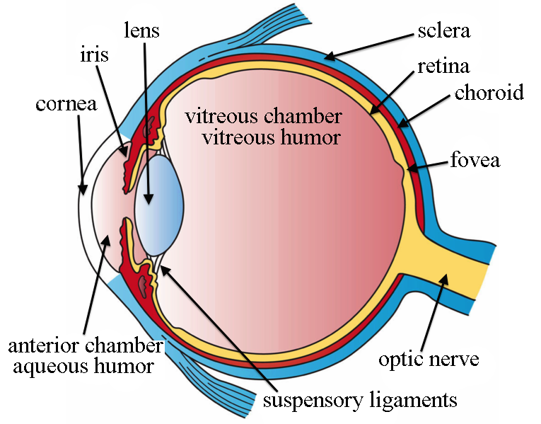
The size of the pupil is controlled by the
A. Ciliary muscles
B. Suspensory ligaments
C. Cornea
D. Iris muscles
Answer
571.5k+ views
Hint: A thin, circular opening in front of the lens is the pupil. It constricts and dilates according to the intensity of light falling on the eyes.
Complete answer:
Eyeball is made up of two segments, an anterior part, and a posterior part. The anterior part is small and forms one-sixth of the eyeball. The posterior part is larger and forms five-sixths of the eyeball. The posterior wall of this part is lined by the light-sensitive structure called the retina. The Wall of the eyeball is composed of three layers:
A. Outer layer, which includes cornea and sclera: Posterior five sixth of this coat is opaque and it is called the sclera. Anterior one-sixth is transparent and is known as the cornea. The cornea is the transparent convex anterior portion of the outer layer of the eyeball, which covers the iris and pupil.
B. Middle layer, which includes choroid, ciliary body, and iris: Choroid is the thin vascular layer of the eyeball situated between the sclera and retina. The ciliary body is the thickened anterior part of the middle layer of the eye, situated between the choroid and iris. Iris is a thin colored curtain-like structure of eyeball, located in front of the lens. It forms a thin circular diaphragm with a circular opening in the center called the pupil. Iris is formed by muscles: i. Constrictor pupillae or iris sphincter muscle or pupillary constrictor muscle: It is formed by circular muscle fibers. The contraction of this muscle causes constriction of the pupil. ii. Dilator pupillae or pupillary dilator muscle: It is formed by radial muscle fibers. The contraction of this muscle causes dilatation of the pupil. Activities of these muscles increase or decrease the diameter of the pupil and regulate the amount of light entering the eye.
C. Inner layer, the retina.

Hence, the correct answer is option (D)
Note: 1.Except anterior one-sixth, the eyeball is situated in a bony cavity known as an orbital cavity or eye socket. Eyeballs are attached to the orbital cavity by the ocular muscles.
2. Suspensory ligaments from the lens are attached to the ciliary body
Complete answer:
Eyeball is made up of two segments, an anterior part, and a posterior part. The anterior part is small and forms one-sixth of the eyeball. The posterior part is larger and forms five-sixths of the eyeball. The posterior wall of this part is lined by the light-sensitive structure called the retina. The Wall of the eyeball is composed of three layers:
A. Outer layer, which includes cornea and sclera: Posterior five sixth of this coat is opaque and it is called the sclera. Anterior one-sixth is transparent and is known as the cornea. The cornea is the transparent convex anterior portion of the outer layer of the eyeball, which covers the iris and pupil.
B. Middle layer, which includes choroid, ciliary body, and iris: Choroid is the thin vascular layer of the eyeball situated between the sclera and retina. The ciliary body is the thickened anterior part of the middle layer of the eye, situated between the choroid and iris. Iris is a thin colored curtain-like structure of eyeball, located in front of the lens. It forms a thin circular diaphragm with a circular opening in the center called the pupil. Iris is formed by muscles: i. Constrictor pupillae or iris sphincter muscle or pupillary constrictor muscle: It is formed by circular muscle fibers. The contraction of this muscle causes constriction of the pupil. ii. Dilator pupillae or pupillary dilator muscle: It is formed by radial muscle fibers. The contraction of this muscle causes dilatation of the pupil. Activities of these muscles increase or decrease the diameter of the pupil and regulate the amount of light entering the eye.
C. Inner layer, the retina.

Hence, the correct answer is option (D)
Note: 1.Except anterior one-sixth, the eyeball is situated in a bony cavity known as an orbital cavity or eye socket. Eyeballs are attached to the orbital cavity by the ocular muscles.
2. Suspensory ligaments from the lens are attached to the ciliary body
Recently Updated Pages
Master Class 11 Computer Science: Engaging Questions & Answers for Success

Master Class 11 Business Studies: Engaging Questions & Answers for Success

Master Class 11 Economics: Engaging Questions & Answers for Success

Master Class 11 English: Engaging Questions & Answers for Success

Master Class 11 Maths: Engaging Questions & Answers for Success

Master Class 11 Biology: Engaging Questions & Answers for Success

Trending doubts
One Metric ton is equal to kg A 10000 B 1000 C 100 class 11 physics CBSE

There are 720 permutations of the digits 1 2 3 4 5 class 11 maths CBSE

Discuss the various forms of bacteria class 11 biology CBSE

Draw a diagram of a plant cell and label at least eight class 11 biology CBSE

State the laws of reflection of light

10 examples of friction in our daily life


