


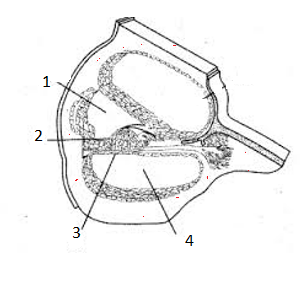
"X" is a spiral-shaped structure (given below) consisting of hair cells that serve as receptors for auditory stimuli. Identify "X" and its label & location (marked as 1, 2, 3, and 4) from the given diagrammatic representation of the sectional view of the cochlea.
A. X- Organ of corti, 2, 3.
B. X- Eustachian tube, 1, 2.
C. X- Semicircular canal, 3, 4.
D. X- Crista ampullaris, 1, 4.
Answer
331.8k+ views
Hint: The innermost ear structure referred to as a cochlea is a snail-shell like structure split into three fluid-filled portions. Two are canals for the transmission of pressure and in the third is the sensitive organ of the Corti, which identifies pressure impulses and will react with electrical impulses which move over the auditory nerve to the brain.
Complete Step-by-step answer: The highlighted labels 1, 2, 3, and 4 are Reissner's membrane, organ of Corti, basilar membrane, and tectorial membrane respectively. The organ of Corti (X) is a sensitive element in the inner ear. It is a spiral-shaped structure situated on the basilar membrane (3) containing hair cells that function as auditory receptors. The hair cells are present in rows on the inner side of the organ of Corti.
Reissner's membrane and the basilar membrane define three fluid-filled chambers within the cochlea such as scala media, scala tympani, and scala vestibuli. The mechanosensitive hair cells live in the organ of Corti on the basilar membrane, which constitutes a single boundary of the scala media.
The basilar membrane is the most important mechanical component of the innermost ear. It has graded mass and stiffness characteristics around its length, and its vibration patterns have the effect of splitting incoming sound into its component frequencies that trigger the various cochlear areas.
The tectorial membrane (TM) of the innermost ear is a ribbon-like strip of the extracellular matrix that spirals along the full length of the cochlea.

Therefore the correct answer is Option A.
Note: The cochlea contains three fluid-filled sections. The perilymph fluid in the canals varies from the endolymph fluid in the cochlear duct. The organ of the Corti is the sensor of the pressure changes.
Complete Step-by-step answer: The highlighted labels 1, 2, 3, and 4 are Reissner's membrane, organ of Corti, basilar membrane, and tectorial membrane respectively. The organ of Corti (X) is a sensitive element in the inner ear. It is a spiral-shaped structure situated on the basilar membrane (3) containing hair cells that function as auditory receptors. The hair cells are present in rows on the inner side of the organ of Corti.
Reissner's membrane and the basilar membrane define three fluid-filled chambers within the cochlea such as scala media, scala tympani, and scala vestibuli. The mechanosensitive hair cells live in the organ of Corti on the basilar membrane, which constitutes a single boundary of the scala media.
The basilar membrane is the most important mechanical component of the innermost ear. It has graded mass and stiffness characteristics around its length, and its vibration patterns have the effect of splitting incoming sound into its component frequencies that trigger the various cochlear areas.
The tectorial membrane (TM) of the innermost ear is a ribbon-like strip of the extracellular matrix that spirals along the full length of the cochlea.
Therefore the correct answer is Option A.
Note: The cochlea contains three fluid-filled sections. The perilymph fluid in the canals varies from the endolymph fluid in the cochlear duct. The organ of the Corti is the sensor of the pressure changes.
Recently Updated Pages
Master Class 8 Social Science: Engaging Questions & Answers for Success
Master Class 8 English: Engaging Questions & Answers for Success
Class 8 Question and Answer - Your Ultimate Solutions Guide
Master Class 8 Maths: Engaging Questions & Answers for Success
Master Class 8 Science: Engaging Questions & Answers for Success
Master Class 7 English: Engaging Questions & Answers for Success
Trending doubts
What are the factors of 100 class 7 maths CBSE
Which are the Top 10 Largest Countries of the World?
What is BLO What is the full form of BLO class 8 social science CBSE
The value of 6 more than 7 is A 1 B 1 C 13 D 13 class 7 maths CBSE
One Metric ton is equal to kg A 10000 B 1000 C 100 class 11 physics CBSE
There are 720 permutations of the digits 1 2 3 4 5 class 11 maths CBSE