


Biology Notes for Chapter 14 Breathing and Exchange of Gases Class 11 - FREE PDF Download
All biological activities require energy to take place and this energy is released by the decomposition of food. So all living things consume food to get high-energy organic molecules that release energy after their decomposition.
The decomposition of food in the presence of oxygen is called oxidation. Cells use oxygen for breaking down complex molecules and produce energy together with oxygen.
Breathing is the process of exchanging atmospheric oxygen with carbon dioxide which is produced by the cells.
The process of oxidation of complex food molecules into simpler molecules within the living cells of an organism is called respiration. The term ‘Respiration’ was coined by Dutrochet. During the oxidation process, the chemical energy stored in the food as complex molecules is released and is temporarily stored in the form of ATP. Breathing is the first step of the respiration process. The molecules of food substances that are decomposed during the respiration process are called respiratory substrates. For example glucose, amino acids, fatty acids, etc.
14.1 Respiratory Organs:
Depending on the body structure and habitat of organisms, the respiratory mechanism is different in different types of organisms and so are the respiratory organs present in them. A table is given below in which organisms and their respiratory organs are given:
14.1.1 Human Respiratory System:
The human respiratory system begins with external nostrils which open into the nasal cavity. The nasal cavity extends into the nasal passage that consists of the larynx, trachea, bronchi, and bronchioles.
The nasal cavity is lined with mucous-secreting cells which secrete a slimy fluid called mucous. It can capture dust and other foreign objects in the inhaled air by keeping the nasal cavity moist. The dust particles present in the air are filtered by the hair present in the nasal cavity. The nasal cavity ends in the beginning part of the pharynx.
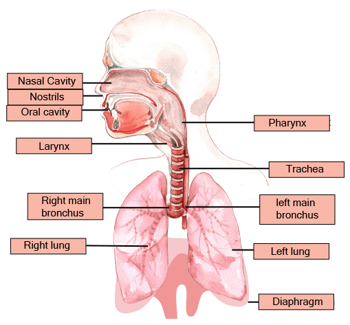
Human Respiratory System
The pharynx is the junction between the respiratory and digestive systems. The oropharynx, Nasopharynx, and Laryngopharynx are the three sections of the pharynx. The oropharynx is the section of the pharynx behind the oral cavity that allows food to pass through, while the nasopharynx is the part that allows air to flow through. The region of the pharynx behind the larynx is known as the laryngopharynx. .The glottis, a slit-shaped aperture at the bottom of the nasopharynx, is protected by the epiglottis, a cartilaginous flap. The main purpose of the epiglottis is to keep food from entering the trachea while swallowing. The glottis opens into the windpipe or trachea, a thin-walled tube that lies in front of the oesophagus and travels through the neck.
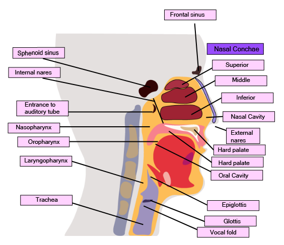
Labeled Diagram of Nasal and Throat Cavity
Labelled Diagram of Nasal and Throat Cavity
The larynx is the enlarged, upper part of the trachea, meant to produce sound. Cartilage rings like thyroid cartilage and cricoid cartilage support the walls of the larynx. In the case of men, the thyroid cartilage is large in size and protrudes from the neck, and is called Adam’s apple. There are two mucous membranes present in the throat cavity and these membranes are called vocal cords. The vocal cords vibrate which in turn produce sounds.
The trachea is protected by C-shaped rings made up of cartilage. When there is less air in the trachea, the wall of the trachea can be prevented from collapsing by these cartilaginous rings.
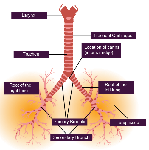
Labeled Diagram of Trachea
Labelled Diagram of Trachea
The trachea is further divided into two bronchi which then enter into the lungs. Each lung has one of these two bronchi. Both of the bronchi divides again to form bronchioles, spread in the entire lungs. Each of the bronchioles is divided into several tiny ducts named alveolar ducts. Each alveolar duct is inflated into thin-walled air sacs called alveoli. Infundibulum is the alveolar group or group of alveoli. Therefore, each of the infundibula seems like a bunch of grapes.
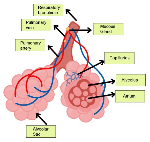
Labeled Diagram of Alveoli
Labelled Diagram of Alveoli
The bronchi, bronchioles, and alveoli are the major components of the lungs. Both of the lungs are protected by a double-layered membrane called the pleura. The pleura is filled with pleural fluid, which reduces friction on the surface of the lungs. The external pleural layer is in contact with the thoracic lining and the internal pleural layer is in contact with the surface of the lung.
The Respiratory System is Divided Into Two Parts:
The Conduction Part: The conduction part comprises all the respiratory components from the nostrils to the alveoli of the lungs. The function of this part is to conduct atmospheric air from external nostrils to the alveoli of the lungs. This part also prevents foreign objects from entering the respiratory passage, it moisturises the transporting air and brings its temperature close to the body temperature.
The Exchange Part: The exchange part consists of the site where Oxygen diffuses between the blood and the atmospheric air present in the lungs.
The lungs are present in the chest cavity. Their dorsal side is bounded by the spine, the ventral side is bounded by the sternum, both of the lateral sides are bounded with ribs, and the bottom with a dome-shaped muscular sheet called the diaphragm.
Any changes in the chest cavity volume are expressed in the lung cavity and this change is necessary for breathing.
The Steps Involved in Breathing and Respiration Processes Are Given Below:
The first step is breathing or lung ventilation (inhalation of atmospheric air and exhalation of alveolar air rich in carbon dioxide.
In the second step, gases(oxygen and carbon dioxide) diffuse through the alveolar membrane
In the next step, diffused gases are transported through the blood to their target organs.
Then diffusion of oxygen and carbon dioxide gases occurs between blood and body tissues.
The body cells use oxygen for catabolic reactions and the resulting release of carbon dioxide gas. This process is also known as cellular respiration.
14.2 Mechanism of Respiration:
The influx and efflux of atmospheric air in the lungs, between the atmosphere and alveoli, is called breathing. It is a consequence of the expansion and contraction of the lungs.
The lungs contract in two processes:
Inspiration or Inhalation
Expiration or Exhalation.
A pressure gradient is created between the lungs and the atmosphere, to accomplish both of the above processes.
Inspiration or Inhalation: It is an active process that means it uses energy to take place and mainly includes muscle contraction. It occurs when the pressure inside the lungs is less than atmospheric pressure. The lungs are located in the chest or thorax. Contraction of the diaphragm (the muscular sheet that divides the chest and abdomen) and some intercostal muscles (muscles present between the ribs) occur during inhalation, as a result, the ribs are pulled upside and in the outward direction. This process results in increasing the volume of the chest cavity because the sternum increases the volume of the thoracic cavity in the dorsal-ventral axis. An overall increase in the volume of the thoracic cavity also increases the volume of the lungs. Increased lung or pulmonary volume reduces the pressure inside the lungs. As a result, fresh air enters the lungs through the nose.
Expiration or Exhalation: Relaxation of the diaphragm and intercostal muscles causes the diaphragm and sternum to return back to their normal positions. This results in a reduction of the thoracic volume (volume of the chest cavity) and pulmonary volume (volume of lungs) but increases the intrapulmonary pressure (pressure inside lungs). This pressure is slightly higher than the atmospheric pressure, which causes the air to be expelled out from the lungs.
The strength of inhalation and exhalation can be increased with the help of extra-abdominal muscles.
\A healthy person normally breathes 12-16 times per minute, which is about 10 litres of air per minute.
The device used to measure respiratory rate is known as a spirometer or respirometer.
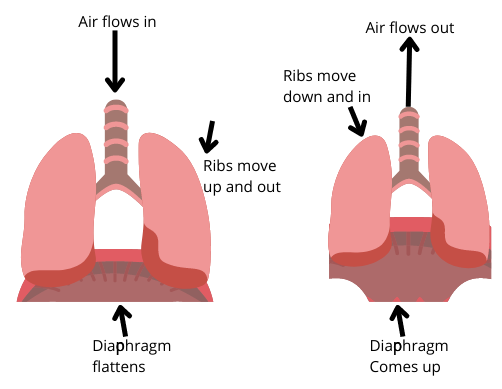
Diagram Showing Mechanism of Respiration
14.2.1 Respiratory Volumes and Capacities:
The air that enters and exits the lungs with each respiration is known as tidal air.
1. Tidal volume (TV): The volume of air inhaled or exhaled during normal breathing is known as the tidal volume. The tidal volume of an adult male is approximately 500 ml. A healthy person can inhale or exhale about 6000-8000 ml of air per minute.
2. Inspiratory reserve volume (IRV): This is the amount of additional air that can be inhaled beyond the normal tidal volume during a forced inhalation. The IRV of a normal person is approximately 2000-3000 ml.
3. Expiratory reserve volume (ERV): This is the additional volume of air that can be exhaled beyond the normal tidal volume during a forced exhalation. The ERV of a normal person is approximately 1100 ml.
4. Residual volume (RV): The residual air volume is the volume of air left in the lungs after forced exhalation. The RV of a normal person is approximately 1100 to 1200 ml. There is always a residual air volume left in the lungs so that even after forced exhalation, gas exchange will continue in the lungs.
5. Pulmonary capacity (PC): The pulmonary capacity is the capacity when two or more lung capacities are considered together.
The important pulmonary capacities of the lungs are given below:
Inspiratory capacity: The total amount of air that a person can inhale after a normal exhalation, is called inspiratory volume. It includes inspiratory reserve and tidal volume.
IC = TV + IRV
3500 mL + 500 mL = 3500 mL
Expiratory Capacity: The amount of air that a person can exhale after a normal inhalation.
EC = TV + ERV
Functional Residual Volume: The amount of air left in the lungs after a normal exhalation. It is the sum total of the expiratory reserve volume and residual volume.
FRC = ERV + RV
2500 ml = 1000 ml + 1500 ml
Vital Capacity: This is the amount of air that is expelled forcefully, after the deepest inhalation. It can be calculated by sum total of inspiratory reserve volume, expiratory reserve volume, and tidal volume.
CV = IRV + ERV + TV
4500 ml = 3000 ml + 500 ml + 1000 ml
Total Lung Volume: The maximum amount of air that the lung can hold is the total lung volume. It is the sum total of inspiratory reserve volume, expiratory reserve volume, tidal volume, and residual volume.
TLC = IRV + ERV + TV + RV or
TLC = VC + RV
14.3 Gas Exchange:
The main part of gas exchange occurs in the alveoli. The exchange of gases takes place between blood and tissues. Gas exchange occurs by simple diffusion based on a pressure gradient or a concentration gradient. The solubility of the gas and the thickness of the film are the important factors affecting diffusion. The partial pressure is the pressure of a single gas in a gas mixture. The partial pressure of oxygen is represented by pO2, and the m=partial pressure of carbon dioxide is represented by pCO2.
Partial pressures of oxygen and carbon dioxide (in mm Hg) at different parts as compared to the other gases present in the atmosphere
Diagrammatic Representation of Gas exchange Between Alveoli and Other Body Parts
The amount of CO2 that can diffuse through the diffusion membrane is greater than the O2 partial pressure. The diffusion membrane is formed by the layer of the squamous epithelium of the alveoli, the endothelium of the capillaries of the alveoli and the basement material between the two layers. The total thickness of the diffusion membrane is less than one millimetre. The diffusion of O2 from the alveoli to the tissues and the diffusion of CO2 from the tissues to the alveoli are aided by all variables in our bodies.
14.4 Gas Transportation:
Both O2 and CO2 gases are transported through the blood. It is reported that 97% of O2 is transported by red blood cells and the remaining 3% is transported through plasma. However, 20-25% of CO2 is transported through RBC, 70% is transported as bicarbonate ions, and the remaining 5-7% of CO2 dissolves in plasma.
14.4.1 Oxygen Transportation:
The red blood cells contain a red-coloured iron-containing pigment that is known as haemoglobin. The iron part of haemoglobin binds with oxygen to form oxyhemoglobin, a combination that is related to the partial pressure of oxygen. One haemoglobin can carry four molecules of oxygen because one molecule of haemoglobin contains four iron-containing parts to which oxygen binds. The binding of oxygen with haemoglobin is affected by CO2 partial pressure, hydrogen ion concentration (pH), and temperature.
The relationship between haemoglobin and oxygen is expressed by plotting per cent saturation of haemoglobin with oxygen against partial pressure of oxygen. This curve is called the oxygen-dissociation curve or oxygen haemoglobin dissociation curve and it is a sigmoid or S-shaped curve.
Oxyhaemoglobin is formed in the alveoli when the pO2 is high, the pCO2 is low, the H+ concentration is low, and the temperature is low. Low pO2, high pCO2, high H+ concentration, and higher temperature occur in the tissues, resulting in oxygen dissociation from oxyhaemoglobin. Hence, oxygen binds to the haemoglobin on the lung surface and is dissociated from the haemoglobin in the tissues. Under normal physiological conditions, 100 mL of oxygenated blood provides about 5mL of oxygen.
14.4.2 Carbon Dioxide Transportation:
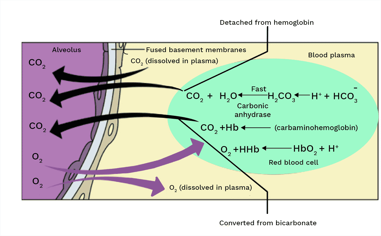
CO2 combines with haemoglobin to form carbamino-haemoglobin, which is proportional to CO2 partial pressure. The binding of carbon dioxide with haemoglobin is affected by the partial pressure of O2. More binding occurs when the partial pressure of CO2 is high and the partial pressure of O2 is low, as more carbon dioxide binding occurs in tissues. When the pCO2 is low and the pO2 is high, as in the alveoli, CO2 dissociates from carbamino-haemoglobin.
The carbonic anhydrase enzyme is abundant in RBCs and is responsible for converting carbon dioxide into bicarbonate ions. This enzyme is only found in trace amounts in plasma.
Because of catabolism, the partial pressure of CO2 in the tissue is high, and it diffuses into the blood to form HCO3- and H+ ions.
CO2 and H2 are formed when the partial pressure of CO2 in the alveolus is low. Bicarbonate ions are formed as a result of carbon dioxide trapping. It moves from the tissue to the alveoli, where carbon dioxide is expelled. 4 mL of carbon dioxide is delivered for every 100 mL of deoxygenated blood.
14.5 Regulation of Respiration Process:
The nervous system regulates the process of respiration or respiratory rhythm. The rate of respiration changes in response to the body's oxygen demand. The respiratory centre is the regulatory centre for respiration. It is made up of a number of neurons located bilaterally in the brain's medulla oblongata. There are three types of respiratory centres: respiratory rhythm centre, pneumatic centre, and chemosensitive area.
14.5.1 Respiratory Rhythm Center:
It is a group of neurons in the medulla oblongata's dorsal region. It is a specialised centre that is in charge of respiration regulation. The basic respiratory rhythm is produced by this group of neurons. Neurons of the respiratory rhythm centre release nervous signals which are transmitted to the diaphragm, the primary inspiratory muscle. These signals cause the inspiratory muscle (diaphragm) to contract, resulting in inspiration.
14.5.2 Pneumotaxic Centre:
It is a group of neurons located dorsally in the brain's upper pons. It has the ability to modulate the functions of the respiratory rhythm centre.
The number and depth of breaths are determined by the signal from the neurons of the pneumatic centre. When the pneumatic centre sends a strong signal, the rate of breathing increases due to the shortening of both inspiration and expiration. The neural signals from the pneumatic centre can also shorten the duration of inspiration and thus change the rate of respiration.
14.5.3 Chemo Sensitive Centre:
This is a region near the respiratory rhythm centre that is extremely sensitive to CO2 and hydrogen ions. The concentration of hydrogen ions and CO2 raises the inspiratory and expiratory signals. There is no direct effect of oxygen on respiratory signals. The chemosensitive centre is activated by the increased concentration of CO2 and Hydrogen ions in the chemosensitive area. Then the signals are transmitted to the respiratory rhythm centre, which adjusts the respiratory process to eliminate these substances.
Changes in the concentration of CO2 and hydrogen ions are recognised by aortic arch and carotid artery receptors. They send the necessary signals to the respiratory rhythm centre in order for immediate control actions to be taken.
14.6 Disorders of the Respiratory System:
Many respiratory diseases and respiratory disorders affect the human respiratory system.
Asthma, emphysema, and occupational respiratory disorders are all common.
Asthma: Asthma is characterised by difficulty breathing and wheezing caused by inflammation of the bronchi and bronchioles.
Emphysema: Emphysema is a chronic obstructive lung disease. It is characterised by abnormal distension or inflation of the alveolar wall, resulting in a loss of elasticity of their walls. Alveolar walls degenerate, and alveoli join together to form large alveoli. Even during expiration, the alveoli remain completely filled with air. This causes an increase in lung size, and cigarette smoking is the leading cause of emphysema.
Occupational Respiratory Disorders: Occupational respiratory disorders are pulmonary diseases caused by exposure to potentially harmful substances such as gas, fumes, or dust in a person's working environment. Silicosis (caused in workers employed in the mining industry, quarry, etc. due to chronic exposure to silica dust from rocks or stones) and asbestosis (caused in workers employed in asbestos factories due to chronic exposure to asbestos dust) are two common examples.
Some Frequently Asked Questions in the Exams from Biology Chapter 14
Section–A (1 Mark Questions)
1. How many molecules of O2 are carried by one haemoglobin?
Ans. Four molecules of O2 are carried by one haemoglobin.
2. What causes the urge of inhalation in humans?
Ans. Rising pCO2 level causes the urge of inhalation in humans.
3. Which enzyme increases the reaction rate between CO2 and H2O in red blood cells?
Ans. Carbonic anhydrase is the enzyme that increases the reaction rate between CO2 and H2O in red blood cells.
4. Write one respiratory disorder.
Ans. Emphysema
5. Which is the prime site for the exchange of gases in the human body?
Ans. Alveoli are the prime site for gaseous exchange in the human body.
Section–B (2 Mark Questions)
6. In an old science fiction movie, the hero tries to drown a giant ant by holding its head underwater. Would this work? Why?
Ans. Ants breathe in oxygen through spiracles which are a series of holes located on the sides of their bodies. Carbon dioxide exiting through said tubes as well. So no, the giant ant cannot die if its head is underwater.
7. A major percentage (97%) of O2 is transported by RBCs in the blood. How is the remaining percentage (3%) of O2 transported?
Ans. Haemoglobin is the Fe-containing respiratory pigment present in blood, and it is a carrier of oxygen. 97% of oxygen is carried with haemoglobin from lungs to tissues and the remaining 3% is dissolved in plasma which carries oxygen to the body cells.
8. Write the organs of respiration in the entities given below:
(i) Flatworm
(ii) Frog
(iii) Birds
(iv) Cockroach
Ans.
(i) Flatworm – Body surface.
(ii) Frog – Moist skin and lungs.
(iii) Birds – Lungs.
(iv) Cockroach – Tracheal tubes
9. Write the name of two parts that are involved in initiating a pressure gradient between the lungs and the atmosphere during normal respiration.
Ans. Diaphragm and a set of external and intercostal muscles between the ribs are involved in the generation of pressure gradient during respiration.
10. Answer the following questions.
(i) What is the amount of O2 supplied to tissues through every 100 mL of oxygenated blood under normal physiological conditions?
(ii) How much CO2 is delivered by 100 mL of deoxygenated blood?
Ans.
(i) Every 100 mL of oxygenated blood can deliver around 5 mL of O2 to the tissues under normal physiological conditions.
(ii) Every 100 mL of deoxygenated blood delivers approximately 4 mL of CO2 to the alveoli.
11. Arrange the following terms based on their volumes in ascending order:
Tidal Volume (TV)
Residual Volume (RV)
Inspiratory Reserve Volume (IRV)
Expiratory Capacity (EC)
Ans. Tidal Volume (500mL) < Residual Volume (1100-1200 mL) < Expiratory capacity (1500-1600 mL) < Inspiratory reserve volume (2500-3000 mL)
5 Important Topics of Biology Class 11 Chapter 14 You Shouldn’t Miss!
Importance of Biology Chapter 14 Breathing and Exchange of Gases Class 11 Notes
Class 11 Biology Chapter Breathing and Exchange of Gases Notes PDF explains how the body takes in oxygen and expels carbon dioxide, which is crucial for survival.
Breathing and Exchange of Gases Short Notes helps students understand the structure and function of the respiratory system, including the lungs, diaphragm, and blood vessels.
The process of breathing and gas exchange is explored, showing how oxygen reaches the cells for energy production.
Students learn how carbon dioxide, a waste product, is removed from the body to maintain healthy bodily functions.
Understanding these concepts is important for grasping how our body’s respiratory system supports overall health.
The notes simplify complex ideas, making it easier for students to study and prepare for exams.
Class 11 Biology Chapter Breathing and Exchange of Gases Notes provides a foundation for further understanding biological processes related to respiration and overall body functions.
The content in the Breathing and Exchange of Gases Short Notes helps students break down and remember key points essential for their biology studies.
Tips for Learning the Class 11 Biology Chapter 14 Breathing and Exchange of Gases
Start by understanding the basic structure of the respiratory system, focusing on the lungs, trachea, and diaphragm.
Learn how the process of breathing works, including inhalation and exhalation, to see how oxygen enters the body and how carbon dioxide is removed.
Focus on how gases are exchanged in the alveoli, the tiny air sacs in the lungs, where oxygen moves into the blood and carbon dioxide is taken out.
Use diagrams to visualise the pathway of air from the nose to the lungs. This helps in remembering the steps involved in breathing.
Understand the role of haemoglobin in carrying oxygen throughout the body, and how it releases carbon dioxide for expulsion.
Practice with simple flow charts that show the process of gas exchange, to make it easier to recall during exams.
Make use of short notes and key points to reinforce your understanding of important terms and concepts in this chapter.
Try answering sample questions related to this topic to test your knowledge and identify areas where more study is needed.
Conclusion
The study of Chapter 14 - Breathing and Exchange of Gases in CBSE Class 11 Biology is crucial for understanding the intricate mechanisms behind respiration in living organisms. This chapter explores various aspects, including the structure and functioning of the respiratory system, the process of breathing, and the exchange of gases in different organisms. By accessing the FREE PDF download of Class 11 Biology Chapter 14 notes, students gain a comprehensive resource to deepen their knowledge and strengthen their grasp on the subject. These notes provide valuable insights into the concepts discussed in the chapter, such as pulmonary ventilation, transport of gases, and regulation of respiration.
Through these notes, students can enhance their understanding of vital topics like respiration in plants, respiratory disorders, and the significance of respiratory pigments. The well-organised content, diagrams, and explanations in the notes aid in simplifying complex concepts, making the learning process more accessible and enjoyable. By studying these Class 11 Biology notes, students can consolidate their understanding of the respiratory system's functioning, appreciate the adaptability of organisms to different environments, and recognise the interdependence between living beings and their surroundings.
Related Study Materials for Class 11 Biology Chapter 14 Breathing and Exchange of Gases
Students can also download additional study materials provided by Vedantu for Biology Class 11, Chapter 14–
Chapter-wise Class 11 Biology Notes PDF Download
Important Study Materials for Class 11 Biology

FAQs on Breathing and Exchange of Gases Class 11 Biology Chapter 14 CBSE Notes - 2025-26
1. What is the fundamental difference between breathing and respiration for a quick summary?
Breathing is the physical process of inhaling oxygen and exhaling carbon dioxide from the lungs. In contrast, cellular respiration is a biochemical process at the cellular level where food (like glucose) is broken down to produce energy (ATP), which requires oxygen and releases carbon dioxide as a waste product. Essentially, breathing facilitates cellular respiration.
2. How can I quickly revise the mechanism of human breathing?
The mechanism involves two main phases driven by pressure gradients created by the diaphragm and intercostal muscles:
- Inspiration (Inhaling): The diaphragm and external intercostal muscles contract, expanding the chest cavity. This increases thoracic volume and decreases pressure inside the lungs, causing atmospheric air to rush in.
- Expiration (Exhaling): The muscles relax, returning the chest cavity to its original size. This decreases thoracic volume and increases pressure inside the lungs, forcing air out.
3. What are the most important respiratory volumes and capacities to remember from this chapter?
For a quick revision, focus on these key concepts as per the CBSE syllabus:
- Tidal Volume (TV): The volume of air breathed during a normal, relaxed breath (approx. 500 mL).
- Inspiratory Reserve Volume (IRV): The additional volume of air that can be forcibly inhaled after a normal inspiration (approx. 2500-3000 mL).
- Expiratory Reserve Volume (ERV): The additional volume of air that can be forcibly exhaled after a normal expiration (approx. 1000-1100 mL).
- Residual Volume (RV): The volume of air remaining in the lungs even after a forceful expiration (approx. 1100-1200 mL).
- Vital Capacity (VC): The maximum volume of air a person can breathe out after a forced inhalation. It is calculated as TV + IRV + ERV.
4. What is the main function of the conducting part versus the exchange part of the respiratory system?
The conducting part, which includes the nostrils, pharynx, trachea, and bronchi, serves to transport atmospheric air to the alveoli. It also cleans the air of foreign particles, humidifies it, and brings it to body temperature. The exchange part, consisting of the alveoli and their ducts, is the actual site where the exchange of oxygen and carbon dioxide occurs between the blood and the inhaled air.
5. Why is the oxygen-haemoglobin dissociation curve sigmoidal (S-shaped)?
The S-shaped curve reflects the cooperative binding of oxygen to haemoglobin. When the first oxygen molecule binds to a haemoglobin subunit, it causes a change in the protein's shape. This change increases the affinity of the other three subunits for oxygen, making it progressively easier for them to bind. This cooperative effect results in a steep slope on the curve, allowing for rapid oxygen loading in the lungs. The plateau at the top indicates saturation.
6. How is the majority of carbon dioxide transported in the blood, and why is this method so effective?
Approximately 70% of carbon dioxide is transported as bicarbonate ions (HCO₃⁻) in the blood plasma. This method is highly effective because it allows large quantities of CO₂ to be transported from tissues to the lungs with minimal change in blood pH. The conversion of CO₂ to bicarbonate is a reversible reaction rapidly catalysed by the enzyme carbonic anhydrase, which is abundant in red blood cells.
7. What key factors influence the binding of oxygen with haemoglobin in the lungs and its release in the tissues?
The binding and release of oxygen are primarily governed by several factors:
- Partial Pressure of O₂ (pO₂): High pO₂ in the lungs promotes the formation of oxyhaemoglobin, while low pO₂ in tissues facilitates its dissociation.
- Partial Pressure of CO₂ (pCO₂): High pCO₂ in tissues lowers haemoglobin's affinity for O₂, causing oxygen release (the Bohr effect).
- H+ Concentration (pH): An increase in H+ ions (lower pH) in active tissues also decreases oxygen affinity, aiding its delivery.
- Temperature: Higher temperatures in metabolically active tissues promote the release of oxygen from haemoglobin.
8. How do the different respiratory centres in the brain work together to regulate breathing?
Breathing regulation is a coordinated effort by centres in the medulla and pons:
- The Respiratory Rhythm Centre in the medulla oblongata generates the primary rhythm for inspiration and expiration.
- The Pneumotaxic Centre in the pons modulates the signals from the rhythm centre, helping to fine-tune the breathing rate and depth.
- A Chemosensitive Area near the rhythm centre is highly sensitive to changes in blood CO₂ and H+ ion levels, signalling the rhythm centre to adjust breathing to maintain homeostasis.
9. What are the critical differences between asthma and emphysema to note for revision?
For quick revision, remember these key distinctions:
- Asthma is an inflammatory condition causing spasms in the bronchi and bronchioles, which leads to wheezing and difficulty breathing. It is often triggered by allergens.
- Emphysema is a chronic disease characterized by the damage and breakdown of alveolar walls. This reduces the surface area available for gas exchange and is most commonly caused by long-term cigarette smoking.
10. What is vital capacity, and what is its physiological significance?
Vital Capacity (VC) is the maximum volume of air a person can exhale after a maximum, forceful inhalation. Its physiological significance lies in indicating the total usable volume of the lungs for gas exchange. A higher vital capacity suggests a stronger respiratory system and better physical fitness, as it allows for a greater volume of air to be exchanged with each breath, which is particularly important during exercise.