

How the Heart’s Chambers and Valves Direct Circulation
The human heart is a vital organ that keeps us alive by pumping blood throughout the body. It works tirelessly to deliver oxygen and nutrients to every cell while carrying away waste products. In this article, we will explore the diagram of the heart, its structure, the heart diagram with labelling, the section of the human heart diagram, and a simple flow chart of heart function. This content is presented in a way that will help students of all levels understand the topic easily.
Structure of the Human Heart
The heart has four chambers:
Upper Chambers (Atria): These include the right atrium and left atrium (also called auricles). They receive blood returning to the heart.
Lower Chambers (Ventricles): These include the right ventricle and left ventricle. They pump blood out of the heart to the lungs or the rest of the body.
Major Blood Vessels
Arteries: Transport oxygenated blood from the heart to various body areas. However, the pulmonary artery is unique; it carries oxygen-poor blood to the lungs.
Veins: Transport blood low in oxygen from the body back to the heart. The pulmonary vein is an exception, carrying oxygen-rich blood from the lungs to the heart.
Layers of the Heart Wall
The heart wall has three layers:
Epicardium (outer layer)
Myocardium (middle layer): This muscular layer is responsible for the heart’s contractions.
Endocardium (inner layer)
Valves of the Heart
The heart contains four valves to prevent backflow of blood:
Aortic Valve: Stops blood from flowing back into the left ventricle from the aorta.
Mitral Valve: Prevents blood returning to the left atrium when the left ventricle contracts.
Pulmonary Valve: Stops blood from flowing back into the right ventricle from the pulmonary artery.
Tricuspid Valve: Prevents blood returning to the right atrium when the right ventricle contracts.
Blood Flow Chart of the Heart
Below is a simple flow chart of heart circulation:
Blood lacking oxygen enters the right atrium from the body through the vena cava.
It then moves into the right ventricle through the tricuspid valve.
The right ventricle pushes this blood to the lungs via the pulmonary artery, where it gets oxygen.
Oxygenated blood travels back to the left atrium from the lungs through the pulmonary vein.
It then passes through the mitral valve into the left ventricle.
The left ventricle pumps this oxygen-rich blood into the aorta, distributing it throughout the body.
Human Heart Diagram (Labelled)
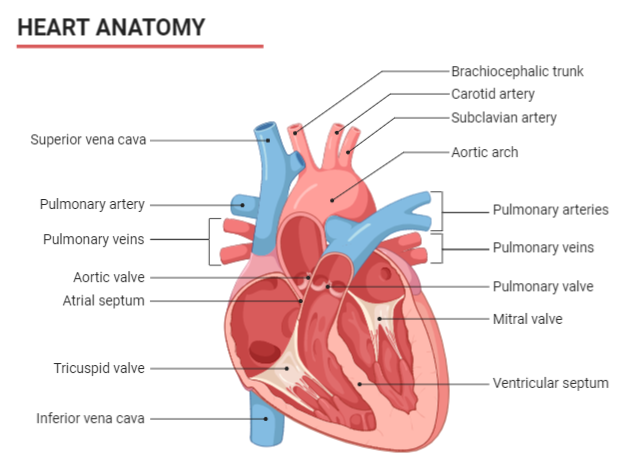
When looking at a human heart diagram, you will notice clear divisions of the atria and ventricles, along with the valves positioned at the boundaries of these chambers. Each component works together to ensure continuous blood circulation.
Quick Quiz
1. Which blood vessel carries oxygenated blood from the lungs to the heart?
Answer: Pulmonary vein
2. Name the valve that prevents backflow of blood into the left ventricle.
Answer: Aortic valve
3. What is the middle layer of the heart wall called?
Answer: Myocardium
4. How many chambers are there in the human heart?
Answer: Four (two atria and two ventricles)
5. Which side of the heart handles deoxygenated blood?
Answer: Right side (right atrium and right ventricle)
Heart Attack Warning Signs
Common warning signs include:
Pain in the chest or discomfort
Shortness of breath
Nausea or vomiting
Excessive sweating
Paying attention to these signs and seeking medical help early can save lives.
Related Topics


FAQs on Human Heart Diagram: Labeled Structure and Blood Flow Explained
1. What are the main chambers and blood vessels shown in a typical heart diagram?
A standard diagram of the human heart illustrates four main chambers and several major blood vessels. The chambers are:
- Right Atrium: Receives deoxygenated blood from the body.
- Right Ventricle: Pumps deoxygenated blood to the lungs.
- Left Atrium: Receives oxygenated blood from the lungs.
- Left Ventricle: Pumps oxygenated blood to the rest of the body.
2. How does blood circulate through the heart? Explain the path of double circulation.
Blood circulates through the heart in a process called double circulation, which involves two distinct pathways: pulmonary and systemic circulation. The path is as follows:
1. Deoxygenated blood from the body enters the right atrium via the vena cava.
2. It flows into the right ventricle, which pumps it into the pulmonary artery.
3. The blood travels to the lungs, releases CO2, and picks up oxygen (pulmonary circulation).
4. Oxygenated blood returns to the left atrium via the pulmonary veins.
5. It then flows into the left ventricle, which pumps it into the aorta.
6. The aorta distributes this oxygen-rich blood to the entire body (systemic circulation).
3. What is the specific function of the four main valves in the heart?
The four heart valves ensure that blood flows in only one direction, preventing any backward leakage. Their specific functions are:
- Tricuspid Valve: Located between the right atrium and right ventricle, it prevents the backflow of blood into the right atrium when the ventricle contracts.
- Pulmonary Valve: Situated between the right ventricle and the pulmonary artery, it stops blood from flowing back into the right ventricle.
- Mitral (Bicuspid) Valve: Found between the left atrium and left ventricle, it prevents blood from returning to the left atrium during ventricular contraction.
- Aortic Valve: Located between the left ventricle and the aorta, it prevents oxygenated blood from flowing back into the left ventricle.
4. Why is the muscular wall of the left ventricle significantly thicker than the right ventricle?
The difference in wall thickness is directly related to the workload of each ventricle. The right ventricle only needs to pump deoxygenated blood a short distance to the lungs, which is a low-pressure circuit. In contrast, the left ventricle must generate much higher pressure to pump oxygenated blood throughout the entire body, from the head to the toes. This requires a stronger, more muscular wall to create the necessary force for systemic circulation.
5. What is the importance of the septum in the heart's structure?
The septum is the muscular wall that divides the left and right sides of the heart. Its primary importance is to prevent the mixing of oxygenated blood (on the left side) with deoxygenated blood (on the right side). This separation is crucial for the efficiency of the circulatory system, ensuring that the body's tissues receive blood with the maximum possible oxygen content. This efficient system supports the high metabolic rate of warm-blooded animals like humans.
6. What is the role of the Sinoatrial (SA) node, and why is it called the 'pacemaker' of the heart?
The Sinoatrial (SA) node is a small cluster of specialised cells located in the upper wall of the right atrium. Its primary role is to generate the electrical impulses that trigger each heartbeat. It is called the natural 'pacemaker' because it sets the rhythm and rate of the heart's contractions. These impulses spread across the atria, causing them to contract, and then travel to the ventricles, ensuring a coordinated and rhythmic pumping of blood.
7. What is the difference between pulmonary circulation and systemic circulation?
Pulmonary and systemic circulation are the two circuits of double circulation, differing in their destination and function:
- Pulmonary Circulation: This is the circuit that moves deoxygenated blood from the right ventricle to the lungs for oxygenation, and then returns the newly oxygenated blood to the left atrium. It is a short, low-pressure system.
- Systemic Circulation: This is the larger circuit that carries oxygenated blood from the left ventricle to all other parts of the body, delivering oxygen and nutrients, and then returns deoxygenated blood to the right atrium. It is a long-distance, high-pressure system.
8. How does the structure of arteries and veins relate to their function in carrying blood?
The structures of arteries and veins are perfectly adapted to their functions:
- Arteries: These vessels carry blood away from the heart under high pressure. Consequently, they have thick, muscular, and elastic walls that can withstand and maintain this pressure, helping to push blood forward.
- Veins: These vessels carry blood towards the heart under low pressure. Their walls are thinner and less elastic. To compensate for the low pressure and prevent the backflow of blood, especially in the limbs, veins are equipped with one-way valves.
9. What is meant by 'cardiac output' and what factors influence it?
Cardiac output (CO) is the total volume of blood pumped by a ventricle of the heart in one minute. It is a critical measure of the heart's efficiency. Cardiac output is calculated by multiplying two key factors:
- Heart Rate (HR): The number of heartbeats per minute.
- Stroke Volume (SV): The volume of blood pumped from a ventricle with each heartbeat.
10. Why is it a misconception that the heart cannot repair itself? What is the actual regenerative capacity of cardiac muscle?
The common belief that the heart cannot repair itself at all is an oversimplification. While it is true that after significant damage, such as a major heart attack, the heart muscle is replaced by non-functional scar tissue, the heart does possess a very limited regenerative capacity. Studies show that cardiac muscle cells (cardiomyocytes) do turn over, but at an extremely slow rate (less than 1% per year). This is insufficient for repairing substantial damage but corrects the misconception that the heart has zero regenerative ability.


