What Are Spores? Importance and Functions in Biology
Spore formation is one of the most reproductive strategies used by a variety of organisms, ranging from fungi and bacteria to algae and plants. Spores are typically single-celled, haploid structures that can develop into new individuals when conditions become favourable. Here, we will explore what is spore, its production, structure and function, and various spore formation examples in diverse living organisms.
What are Spores?
In biology, spores can be defined as specialised reproductive cells formed as a result of either sexual or asexual reproduction. Unlike gametes, spores do not need to fuse with another cell to develop into a new organism. This means they can initiate growth independently, making them crucial for survival and dispersal in many species.
Spore Formation Short Definition
Spore formation refers to the biological process in which spores are produced by certain organisms. Formally known as sporogenesis, this process allows the organism to endure unfavourable conditions and propagate once the environment becomes suitable again.
Structure of Spore (Spore Structure and Function)

A typical spore is a tiny, usually microscopic, unicellular body. While spore structure can vary across different organisms, most spores include:
Protective Coat: A tough outer layer that shields the spore from harsh conditions such as extreme temperatures and radiation.
Cytoplasm and Organelles: Containing essential cellular machinery required to kick-start growth.
Genetic Material (Nucleus): Usually in a haploid state (n), carrying the hereditary information needed to form a new organism.
The spore structure and function together ensure that these cells can remain dormant for extended periods and later germinate into fully functioning organisms once conditions improve.
Spore Formation in Different Organisms
1. Fungi
Many fungi produce spores on specialised structures known as sporangia. For instance, in species like Aspergillus and Penicillium, the sporangium contains numerous minute haploid spores. When these spores are released and land in favourable conditions, they germinate into new fungal hyphae.
Spore Formation Examples in Fungi:
Aspergillus
Penicillium
Rhizopus (detailed below)
2. Bacteria
Certain bacteria form endospores, which are highly resistant structures produced during unfavourable conditions such as nutrient depletion or extreme environmental changes. These endospores are not considered “true” reproductive spores because their primary function is survival rather than reproduction.
Common Bacterial Endospore Examples:
Bacillus species
Clostridium species
For more details on bacterial sporulation, check out Endospores.
3. Plants
In many lower plants (e.g., liverworts, mosses, hornworts), what are spores in plants? They are asexual reproductive units formed in the diploid sporophyte stage. These haploid spores develop into a new generation known as the gametophyte, which eventually produces gametes.
This cycle, known as the alternation of generations, ensures genetic diversity and helps plants adapt to various environmental conditions.
Spore Formation in Rhizopus

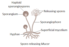
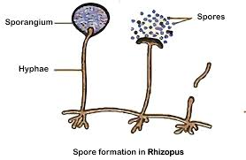
Spore formation in Rhizopus (a common bread mould) is a classic example often studied in schools. Rhizopus produces spores in round sporangia located at the tips of upright hyphae called sporangiophores.
Sporangiophore Formation: The fungus develops upright hyphae, each bearing a bulb-like structure (sporangium) at its tip.
Spore Production: Within each sporangium, several tiny, haploid spores form through asexual reproduction.
Spore Release: When the sporangium matures, it bursts, releasing spores into the environment.
Germination: If spores land in a favourable setting (optimal temperature, moisture, and nutrients), they germinate and grow into new fungal hyphae.
Spore Formation Diagram (Conceptual)
Hypha (mycelium)
|
| (upright growth)
v
Sporangiophore
|
| (knob-like swelling)
v
Sporangium
|
| (contains numerous spores)
v
Burst -> Spores Released -> Germination -> New Fungal Colony
This simple flowchart helps visualise how spores are formed and released in Rhizopus and other fungi.
Additional Insights
Spore Versus Gamete: Spores can develop directly into a new individual, whereas gametes (like sperm and egg) must fuse for fertilisation.
Importance of Spores: They help in dispersal, survival under harsh conditions, and colonising new environments.
Evolutionary Advantage: The ability to remain dormant for extended periods and later germinate ensures the continuity of the species.
Short Quiz (With Answers)
1. Question: Which structure in fungi typically produces spores?
Answer: The sporangium.
2. Question: Name one bacterial genus that forms endospores.
Answer: Bacillus (e.g., Bacillus subtilis).
3. Question: What is the main function of spores in plants?
Answer: To ensure asexual reproduction and facilitate alternation of generations.
4. Question: In spore formation in Rhizopus, what is the name of the structure that holds the spores before they are released?
Answer: The sporangium is located on a sporangiophore.
Conclusion
Spore formation is a vital process in what is spore in biology. It ensures the survival, dispersal, and reproduction of diverse organisms, from fungi and bacteria to algae and plants. By forming spores, these organisms can withstand unfavourable conditions and flourish when the environment becomes supportive.
Related Topics at Vedantu


FAQs on Spore Formation Explained: Structure, Types, and Examples
1. What is spore formation and which organisms use this method of reproduction?
Spore formation is a type of asexual reproduction where a parent organism produces numerous, tiny reproductive units called spores. These spores can develop into new individuals under favourable conditions. It is commonly observed in non-flowering plants like ferns and mosses, as well as in fungi such as Rhizopus (bread mould) and bacteria.
2. What is the typical structure of a spore and how does it help in survival?
A spore is a single-celled reproductive unit with a simple structure designed for survival and dispersal. Its key components are:
- A hard, protective coat: This thick outer wall shields the spore from harsh environmental conditions like extreme temperatures, dehydration, and lack of nutrients.
- Cytoplasm and Nucleus: Inside the coat, the spore contains the cytoplasm and a nucleus with the complete genetic material of the parent.
- Minimal Stored Food: It contains a small food reserve to sustain it until germination begins in a suitable environment.
3. What are the structures that produce and hold spores called?
The structure responsible for producing and containing spores is called a sporangium (plural: sporangia). In common fungi like bread mould (Rhizopus), the sporangium is a round, bulbous structure located at the tip of a stalk-like hypha called a sporangiophore. When mature, the sporangium ruptures, releasing the spores into the surroundings.
4. How do bacterial endospores differ from the reproductive spores of fungi?
Bacterial endospores and fungal spores serve different primary functions. Fungal spores are created for reproduction, aiming to multiply and create new organisms. In contrast, bacterial endospores are dormant, non-reproductive structures formed for survival. A bacterium forms an endospore to protect its DNA during extreme stress and germinates back into a single active cell when conditions improve, not to increase its population.
5. What are the main environmental advantages of reproducing through spores?
Spore formation offers several significant advantages for an organism's survival and propagation:
- High Durability: The tough outer coat allows spores to remain dormant and survive in harsh conditions like extreme heat, cold, and drought for extended periods.
- Widespread Dispersal: Spores are lightweight and produced in massive quantities, enabling them to be easily dispersed over long distances by wind, water, or animals to colonise new areas.
- Rapid Colonisation: Releasing a large number of spores at once increases the probability that some will land in a suitable location and germinate quickly, leading to rapid population growth.
6. How does spore formation fundamentally differ from budding?
While both are forms of asexual reproduction, they are distinct processes. In budding, a new individual grows as a small outgrowth (a bud) directly from the parent's body, as seen in yeast and Hydra. This bud eventually detaches to become a new organism. In spore formation, the parent organism releases numerous specialised, single-celled spores that are typically dormant and can withstand harsh conditions before developing into new individuals. Budding involves direct growth, whereas spore formation involves a dormant, dispersal stage.
7. Do asexually produced spores have any genetic diversity?
Generally, spores produced through asexual means are genetically identical to the parent organism because they are formed via mitosis. This results in very limited genetic diversity. However, many organisms that use spore formation, such as fungi, are also capable of sexual reproduction. This sexual process produces different types of spores that are genetically diverse, as they combine genetic material from two parents.
8. What are the key differences between a spore and a seed?
No, spores and seeds are not the same. They differ in complexity, origin, and resources. Key differences include:
- Structure: A spore is a simple, unicellular (single-celled) body. A seed is a complex, multicellular structure containing a developed embryo, a food supply (endosperm), and a protective seed coat.
- Origin: Spores are typically a product of asexual reproduction in organisms like fungi. Seeds are the result of sexual reproduction (fertilisation of an ovule) in flowering and cone-bearing plants.
- Food Reserve: Seeds possess a substantial food reserve to nourish the embryo during germination. Spores have very minimal food reserves, just enough to survive until they find a suitable substrate to grow on.


