Courses
Courses for Kids
Free study material
Offline Centres
More
Store Icon
Store

Important Questions for CBSE Class 11 Biology Chapter 17 - Locomotion and Movement

ffImage
widget title icon
Latest Updates

widget icon
Enrol Now :
NEET Test Series
widget icon
Grab Your Seat :
NEET Pro Course
widget icon
Register Today :
NEET One to One Coaching

CBSE Class 11 Biology Chapter-17 Important Questions - Free PDF Download

Free PDF download of Important Questions with Answers for CBSE Class 11 Biology Chapter 17 - Locomotion and Movement prepared by expert Biology teachers from latest edition of CBSE(NCERT) books. Register online for Biology tuition on Vedantu.com to score more marks in your examination.


Download CBSE Class 11 Biology Important Questions 2024-25 PDF

Also, check CBSE Class 11 Biology Important Questions for other chapters:

Competitive Exams after 12th Science
tp-imag
bottom-arrow
tp-imag
bottom-arrow
tp-imag
bottom-arrow
tp-imag
bottom-arrow
tp-imag
bottom-arrow
tp-imag
bottom-arrow

Study Important Questions for Class 11 Biology Chapter 17 – Locomotion and Movement

1 Mark Questions:

1. Name the functional contractile unit of a muscle. 

Ans: The functional contractile unit of a muscle is known as a sarcomere.


2. What is arthritis? 

Ans: Arthritis is a condition that causes inflammation of one or more joints, pain and stiffness that can worsen with age.


3. What is the total member of bones present in the left pectoral girdle and the left arm respectively in a normal human? 

Ans: The total number of bones present in the left pectoral girdle is 2 (the clavicle and scapula). Left-arm possesses 30 bones in a normal human.


4. Name the tissue which connects muscles to the bone? 

Ans: Tendon is a fibrous connective tissue that connects muscles to the bones. 


5. What is the function of myoglobin? 

Ans: The function of myoglobin is to store oxygen in the muscles. 


6. What causes fatigue of muscle fibres? 

Ans: Accumulation of lactic acid causes fatigue of muscle fibres.


7. What is a tendon? 

Ans: The tendon is a tough non-elastic connective tissue that joins muscle to a bone.


8. Which type of movable joint makes the hip joint? 

Ans: Ball and socket joint.


9. Name the heaviest and longest bone in the human body? 

Ans: The femur is the heaviest and longest bone in the human body. 


10. How many bones are present in each limb? 

Ans: 30 bones are present in each limb (the upper limb and the lower limb).


11. Why do skeletal muscles show striation? 

Ans: Skeletal muscles show striation due to the distribution pattern of actin and myosin protein (contractile proteins). 


12. Name the last two pairs of ribs. 

Ans: The last two pairs of ribs (11th and 12th Pairs) is known as floating ribs.


13. What lubricates the freely movable joints at the shoulder? 

Ans: The freely mobile joints of the shoulder are lubricated by synovial fluid (synovia).


14. Name of the longest bone of the human body. 

Ans: The femur also called the thigh bone is the longest bone of the human body.


15. Give the name of the first vertebra. 

Ans: The name of the first vertebra is Atlas.


16. Define a sarcomere. 

Ans: Sarcomere, the functional unit of contraction, is the portion of the myofibril between two successive Z lines.


17. Name the cup-shaped bone that constitutes the knee cap. 

Ans: A cup-shaped bone called the patella constitutes the knee cap.


2 Mark Questions:

1. List functions of the skeleton in higher animals? 

Ans: Functions of the skeleton in higher animals are as follows: - 

(i) The skeleton forms the framework/structure of the body.

(ii) The bones of the skeletal system protect the delicate internal organs of the body.

(iii) The skeleton serves as an attachment surface for the body's muscles, tendons, and other similar structures, thus helping in movement.

(iv) It gives the body form and posture.

(v) It helps in the formation of certain blood cells such as RBCs and WBCs.

(vi) It stores minerals such as calcium and phosphorous releases into blood whenever needed.


2. Define a joint. 

Ans: A joint is the part of the body at which two or more bones articulate to allow movement. The surfaces of the two bones are opposite to each other at the joints. These joints assist in the movement of bones in several different ways. The six types of freely movable joints are- ball and socket joint, saddle joint, pivot joint, hinge joint, condyloid joint, and gliding joint.


3. What is osteoporosis? Name two factors that are responsible for osteoporosis. 

Ans: Osteoporosis (porous bone) is an age-dependent systemic bone disorder, characterized by low bone mass and bone mineral density. Factors responsible for osteoporosis are – 

(i) Deficiency of calcium and vitamin D.

(ii) Imbalance of hormones like a parathyroid hormone, thyrocalcitonin and sex hormones.


4. Which kinds of muscle fibres are richly found in the extensor muscles present on the back of the human body? What characteristics enable those fibres to serve their purpose? 

Ans: Extensor muscles on the back of the human body have a high concentration of red muscle fibres. The following are features of red muscle fibres that allow it to perform its function:-

(i) They are rich in mitochondria and myoglobin.

(ii) They carry out a slow rate of contractions for a prolonged time, without getting fatigued.

(iii) No accumulation of lactic acid. fibre

(iv) They generate energy (ATP) by the aerobic catabolism of glucose.


5. Give differences between red and white muscle fibres, other than colour. 

Ans: The difference between red and white muscle fibres are:

S.No

Red Muscle Fibre

White Muscle Fibre


Red muscle fibres are thinner and smaller than white muscle fibres.

White muscle fibres are thicker and larger than red muscle fibres. 


They possess large amounts of myoglobin. 

They possess small amounts of myoglobin. 


They have mitochondria in large numbers. 

They have a comparatively smaller number of mitochondria. 


They carry out a slow rate of contractions for a long period. 

They carry out a fast rate of contraction for a short duration. 


They provide energy by performing aerobic respiration.

They provide energy by performing anaerobic respiration.


6. What are floating ribs? How many of them are there? 

Ans: The last two pairs of ribs, i.e., the 11th and 12th pairs are known as the floating ribs. The floating ribs are smaller in size and have cartilaginous tips. They are only attached dorsally to the respective thoracic vertebrae, so they are also known as the vertebral ribs, and are free ventrally, hence the name "floating ribs". 


7. Why can a red muscle fiber work for a prolonged period, while a white muscle fibre suffers from fatigue soon? 

Ans: 

Red muscle fibres work for a prolonged period because:

(i) They are thin and small muscle fibres.

(ii) They possess large amounts of myoglobin. 

(iii) They have mitochondria in large numbers. 

(iv) They provide energy by performing aerobic respiration.

(v) They carry out a slow rate of contractions for a long period.

White muscle fibres suffer from fatigue soon because:

(i) They are thick and large muscle fibres.

(ii) They possess small amounts of myoglobin. 

(iii) They have a comparatively smaller number of mitochondria. 

(iv) They provide energy by performing anaerobic respiration.

(v) They carry out a fast rate of contraction for a short duration. 


8. What is the function of girdles? 

Ans: There are two girdles in the body, the pectoral girdle and the pelvic girdle. 

A. The function of pectoral girdle:

  • It helps in the articulation of the upper limbs with the axial skeleton. It is composed of two bones: - Clavicle or collar bones and scapula or shoulder bone.

  • It supports your shoulder, facilitates a full range of motion, and protects the nerves and blood vessels that run from the spine to the upper limbs.

  • It performs functions like lifting, holding etc.

B. The function of pelvic girdle:

  • It helps in the articulation of the lower limbs with the axial skeleton.  It is composed of three bones: - the upper ileum, inner pubic, and ischium.

  •  It helps in carrying the weight of the body. 

  • It supports and balances the trunk and the intestines, urinary bladder, and internal sex organs.

  • It performs functions like walking, standing, jumping, running etc.


9. What makes the synovial joints freely movable? List any four types of synovial joints.

Ans: Synovial Joints: - In the synovial joints, a space called the synovial cavity is present between articulating bones. This cavity is filled with lubricating synovial fluid, which reduces the friction on the articulating surface of bones and helps to provide extra cushioning against impact. Hence, the synovial joints are freely movable.

Synovial joints are of the following types: 

(i) Ball and Socket Joint: - Ball and socket joint is present in between the head of the femur and acetabulum.

(ii) Hinge Joint: Hinge joint is present in between phalanges.

(iii) Pivot Joint: Pivot joint is present in between the Atlas and Axis.

(iv) Saddle Joint: The saddle joint is located between the carpal and metacarpal of the thumb.


3 Mark Questions:

1. Explain the initiation of muscle contraction. What is the role of the sarcoplasmic reticulum, myosin head and F-actin during contraction in striated muscles?

Ans: Initiation of muscle contraction: - Muscle contraction is initiated by a signal that is sent by a CNS (Central nervous system) through a motor neuron. A neuromuscular junction or motor-end plate is a junction between a motor neuron and the sarcolemma of the muscle fibre. When a signal reaches the neuromuscular junctions, Acetylcholine (a neurotransmitter) is released which results in the generation of an action potential in the sarcolemma. This spreads through the muscle fibres, resulting in the release of calcium ions from the sarcoplasmic reticulum into the sarcoplasm. Calcium ions bind with the subunit of troponin on actin filaments and thus remove the masking of active sites for myosin. Hence, active sites on actin are exposed and this allows myosin heads to attach to this site. 


Initiation of Muscle Contraction

Initiation of Muscle Contraction


Sarcoplasmic Reticulum: It releases calcium ions into the sarcoplasm that bind with a subunit of troponin on actin and brings about conformational changes. So, they remove the masking of the active binding sites for myosin on the actin filaments. 

Myosin Head: It is an active ATPase enzyme. It provides specific binding sites for ATP and active sites for F-actin to form cross-bridges.

F-Actin: The active binding sites on F-actin are specific to the myosin head and are required for cross-bridge formation.


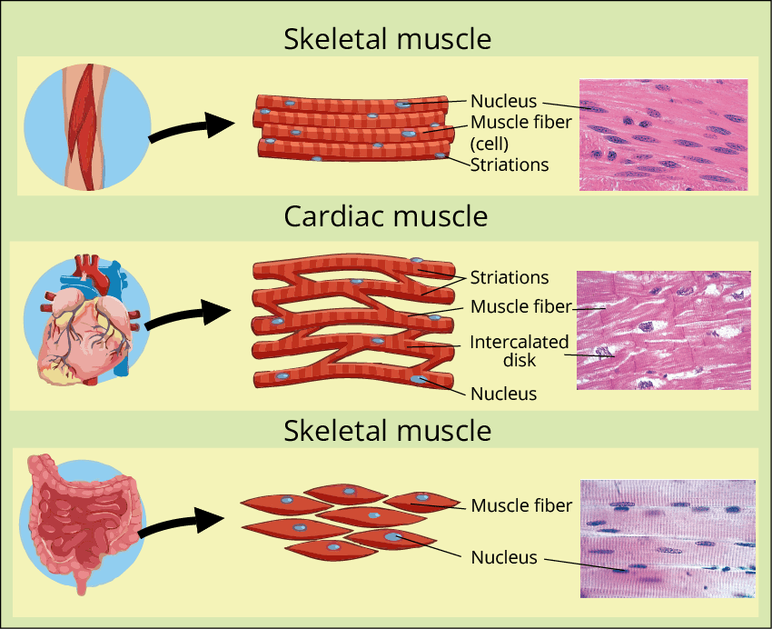
2. What are the three types of muscle tissue? Write two characteristic points about the structure of each of them?

Ans: Types of muscle tissue: - Muscle tissue is of three types- Skeletal, visceral (smooth) and cardiac.

(i) Skeletal/Striated Muscles:

  • The cells of the skeletal muscles are straight, cylindrical, non-branched and multinucleated. They show prominent striations and are thus called striated.

  • They are voluntary muscles as their activities are under the voluntary control of the nervous system.

(ii) Visceral/Smooth/Non-Striated Muscles:

  • The cells of the smooth muscles are narrow, spindle-shaped and uni-nucleated. They do not have any striation and are smooth in appearance, hence called smooth or non-striated muscle.

  • They are involuntary muscles as their activities are not directly controlled by the nervous system. They are controlled by the nervous system, endocrine system and different chemicals.

(iii) Cardiac Muscles:

  • The cells of the cardiac muscles are cylindrical, branched and uni-nucleated. They are striated. 

  • They are involuntary muscles as their activities are not directly controlled by the nervous system. They are controlled by the central nervous system, endocrine system and different chemicals.


Type of Muscles

Type of Muscles


3. Represent diagrammatically a sarcomere and label its parts. Which of these parts shorten during muscle contraction?

Ans:


Sarcomere

Sarcomere


Each muscle fibre consists of alternate light and dark bands. Myosin is a thick contractile protein found in the dark band and is known as the A-band or Anisotropic band. Actin is a thin contractile protein found in the light band and is known as the I-band or Isotropic band.  Each I-band is bisected by an elastic fibre known as the Z line. The thin filament (actin) is firmly attached to the Z line. The H-zone is the central portion of the thick filament (myosin) that is not overlapped by the thin filament. Sarcomere, the functional unit of contraction, is the portion of the myofibril between two successive Z lines.

During muscle contraction, the myosin heads come into close contact with the thin filaments. As a result, the thin filaments are pulled towards the centre of the sarcomere as well as the Z line attached to the actin filaments is also pulled, causing shortening of the sarcomere. The length of the A-band or anisotropic band remains constant as its original length whereas the I-band or isotropic band shortens and the H-zone disappears.


4. Describe any three disorders of the muscular system.

Ans: Three disorders of the muscular system are 

(i) Myasthenia Gravis: This is a type of autoimmune disease. A breakdown in communication between nerves and muscles causes this condition. It affects the neuromuscular junction that causes progressive weakening and paralysis of skeletal muscles. Symptoms are double vision, weakness in the arm and leg muscles, and difficulties with speech and chewing. 

(ii) Muscular Dystrophy: It is a genetic disorder that causes weakness and progressive degeneration of skeletal muscles. Symptoms include trouble breathing and swallowing.

(iii) Tetany: It refers to the rapid spasm (wild contraction) or the continued state of contraction due to low Ca++ in the body fluid and hyperparathyroidism. Symptoms include muscle spasms, speaking and breathing difficulty, numbness in hands and feet, seizures and heart problems.


5. Differentiate between Endoskeleton and Exoskeleton.

Ans: The difference between endoskeleton and exoskeleton are: 


Endoskeleton

Exoskeleton


The endoskeleton (internal skeleton) of an animal is present inside the body.

The exoskeleton (external skeleton) of an animal is present outside the body.


It is made up of hard bones and cartilages.

It is made up of cuticles, scales, and shells made up of chitinous tissues.


It develops from the endoderm.

It develops from the ectoderm.


It is found in vertebrates, namely amphibians, reptiles, fishes, birds and mammals.

It is found in invertebrates such as insects, spiders, centipedes, millipedes, crabs etc.


6. Explain the following-

  1. Antagonistic Muscles

Ans: Contraction of muscles that results in the opposite movements at the same joint is called antagonistic muscles, i.e. When one muscle contracts, the other of the muscle undergoes relaxation and vice versa. Examples include biceps and triceps, quadriceps and hamstrings. During flexion at the elbow, the biceps contracts and triceps relaxes whereas during extension triceps contracts and biceps relaxes.

  1. Tetanus

Ans: If a muscle fibre is stimulated by many nerve impulses or electric shocks it will remain in the state of contraction till the stimulation continues is known as tetanus. It refers to the rapid spasm (wild contraction) or the continued state of contraction due to low Ca++ in the body fluid and hyperparathyroidism. Symptoms include muscle spasms, speaking and breathing difficulty, numbness in hands and feet, seizures and heart problems.

  1. Threshold Stimulus

Ans: Each skeletal muscle is made up of many muscle fibres, each of which is supplied by a nerve. These nerves transmit nerve impulses to muscle fibres. As a result, the muscle is stimulated, and contraction occurs. However, contraction of muscle fibres necessarily requires a minimum strength of the nerve impulse. This is called threshold stimulus.


5 Mark Questions:

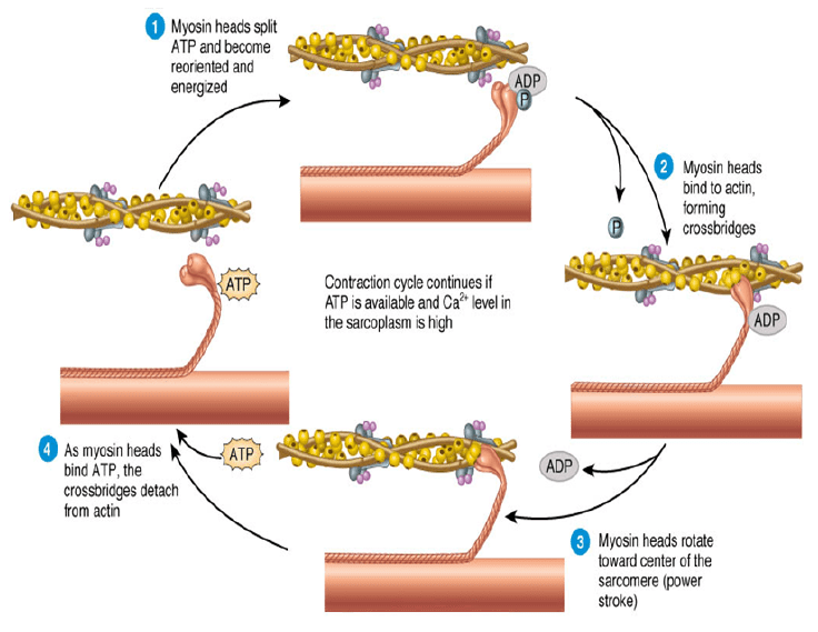
1. What is the role of Ca++ and ATP in muscle contraction? 

Ans: Role of Ca++ in muscle contraction: - During skeletal muscle contraction, the thick filament slides (myosin) past the thin filament (actin) via repeated binding, releasing myosin along the filament.  Muscle contraction is initiated by a signal that is sent by a CNS (Central nervous system) through a motor neuron. When a signal reaches the neuromuscular junctions, Acetylcholine (a neurotransmitter) is released which results in the generation of an action potential in the sarcolemma. This spreads through the muscle fibres, resulting in the release of calcium ions from the sarcoplasmic reticulum into the sarcoplasm. Calcium ions bind with the subunit of troponin on actin filaments and thus remove the masking of active sites for myosin. 

Role of ATP in muscle contraction: - Adenosine triphosphate (ATP) is required for muscular contraction. When ATP binds to the myosin head, it causes the cross-bridge between actin and myosin to detach. ATPase is the enzyme that is found at the binding site on myosin. ATP supplies the energy to pull the myosin head back by hydrolyzing to ADP and inorganic phosphate (Pi), preparing for the next cycle.


Muscle Contraction

Muscle Contraction


2. Describe the various kinds of joints in the human body. According to mobility, give one example of each. 

Ans: A joint is the part of the body at which two or more bones articulate to allow movement. The surfaces of the two bones are opposite to each other at the joints. These joints assist in the movement of bones in a number of different ways. Three types of joints are found in the body of vertebrates. These are:

A. Perfect Joints: The perfect joints have synovial joints and can perform movements in more than one plane. They are of the following 6 types:

(i) Ball and Socket Joints: As the name implies, one bone forms a cup-like depression of socket in which a ball-like structure fits. The head or ball of the joint can freely move in any direction. Example: Shoulder and hip joints. 

(ii) Hinge Joint: In this type of joint, movement can only be performed in one direction. Example: Elbow joint, knee joint, joints of phalanges of fingers and toes. 

(iii) Gliding Joints: It enables bones to glide past one another in any direction along the plane of a joint, i.e., from left and right, up and down, and diagonally. Such joints are found in the vertebral column and the bones of the wrist and ankles.

(iv) Pivot Joint: One bone of this joint is always fixed, while the other can freely move over the former. Example: Neck Joint.

(v) Saddle Joints: It is a biaxial joint that allows movement on two planes i.e., flexion/extension and abduction/adduction. Example: Thumb.

(v) Condyloid Joints: These are joints with two-axis that allow side to side and up to down motions. Example: Base of the index finger and carpals of the wrist.

B. Imperfect Joint: Imperfect joints are the joints that do not possess synovial capsules or connecting ligaments. Example:  The joints between the ilium of the pelvic girdle and the transverse process by sacral vertebra.

C. Immovable Joints: Joints that are permanently fixed and cannot perform any movement are termed immovable joints. These also do not possess synovial capsules of ligaments and do not allow any kind of movement. Example: Bones of the skull and pelvic girdle.


3. Explain the sliding filament theory of muscle contraction.

Ans. The sliding filament theory describes the process of muscle contraction in which the thick filaments (myosin) of muscle fibres slide past the thin filaments (actin), resulting in the shortening of the myofibril. Each muscle fibre consists of alternate light and dark bands. They contain a unique contractile protein called actin and myosin respectively. Myosin is a thick contractile protein found in the dark band and is known as the A-band or Anisotropic band. Actin is a thin contractile protein found in the light band and is known as the I-band or Isotropic band.  Each I-band is bisected by an elastic fibre known as the Z line. The thin filament (actin) is firmly attached to the Z line. The H-zone is the central portion of the thick filament (myosin) that is not overlapped by the thin filament. Sarcomere, the functional unit of contraction, is the portion of the myofibril between two successive Z lines.

During muscle contraction, the myosin heads come into close contact with the thin filaments. As a result, the thin filaments are pulled towards the centre of the sarcomere as well as the Z line attached to the actin filaments is also pulled, causing shortening of the sarcomere. The A-band, or anisotropic band, stays the same length as before, whereas the I-band, or isotropic band, shortens and the H-zone vanishes.


Sliding Filament Theory

Sliding Filament Theory


Important Related Links for CBSE Class 11 

FAQs on Important Questions for CBSE Class 11 Biology Chapter 17 - Locomotion and Movement

1. Why is Chapter 17 - Locomotion and Movement of Class 11 Biology important?

Except for plants, which are immobile, living organisms display locomotion and movement as one of their characteristics. Students would have learned the fundamentals of locomotion and movement in earlier years. In Class 11, students learn more about the mechanics of movement and other related topics as they delve further into the subject. One of the most important characteristics of living things is movement. Plants and animals may move in a variety of ways. Locomotion is the voluntary movement of a person. Organisms migrate for a variety of reasons, including food, refuge, and avoiding predators. The modes of movement used by animals differ.

2. Are NCERT Solutions enough to prepare Chapter 17 - Locomotion and Movement of Class 11 Biology?

The material of the NCERT Solutions for Chapter 17 - Locomotion and Movement of Class 11 Biology is based on the textbook. The topics are taught in an easy-to-understand manner such that students can learn things easily. Wherever possible, analogies are utilised to help students prepare for their second-term exams. Keeping visual learning in mind, neatly labelled graphics for each subject are developed. Tables and points are utilised wherever appropriate to assist pupils in quickly grasping new ideas. The NCERT Solutions not only assist students with their second-term exam preparation, but they also boost their confidence to take competitive examinations such as JEE, NEET, and others.

3. What is Chapter 17 - Locomotion and Movement of Class 11 Biology about?

Locomotion and Movement is one of the chapters of Unit 5- Human Physiology, in the NCERT Biology for Class 11. The chapter discusses the types of movement that occur in both the human body and unicellular creatures. This chapter covers a variety of muscle motions, joint movements, and the skeletal structure of the body, among other essential subjects. The students will also learn about skeletal and muscular system diseases in this chapter. The use of colourful graphics piques students' attention and makes studying more enjoyable.

4. What is the weightage of  Chapter 17 - Locomotion and Movement of Class 11 Biology?

The fifth unit is divided into seven chapters. The 20th chapter of the CBSE Biology syllabus for Class 11 is Locomotion and Movements. The fifth unit is one of the most significant, since it accounts for 18 points in final exams. Every chapter in this unit has a high chance of receiving the highest possible grade. Furthermore, because the NEET examinations provide a 20% weightage to these five units, students should begin studying the NCERT Solutions for Class 11 Biology Locomotion and Movement as soon as possible.

5. What is the importance of NCERT Solutions for Chapter 17 - Locomotion and Movement of Class 11 Biology?

NCERT Solutions have covered all the concepts of the Chapter 17 - Locomotion and Movement of Class 11 Biology in order to assist students in achieving excellent outcomes in their exams. The chapter's exact overview aids students in reviewing each topic before exams. Biology professors with years of experience are the ones that provide all of the correct answers with a variety of examples. Students may readily comprehend the bodily anatomy when given a diagrammatic description. Students may be confident that if they use the Locomotion and Movement Class 11 NCERT Solutions, they will receive outstanding grades and obtain the best possible scores on their exams.