


Locomotion and Movement: An Introduction
Movement is the point at which the living organism moves a body part or parts to bring without a change of the place of the organism. Locomotion is the point at which the movement of a part of the body prompts a change in the position and area of the life form. Both of these are achieved by the joint efforts of the skeletal and muscular systems working closely together. Movement is seen in both vertebrates and non-vertebrates.
Movement is the change in body posture whereas locomotion is the change in place or location. Locomotion is generally performed for food, shelter, mate, specific breeding ground and favourable climatic conditions.
What are Locomotion and Movement?
Locomotion is the voluntary movement of a person starting with one spot and then onto the next. Strolling, running, climbing, and swimming are the instances of motion. All locomotion is movement, however all movements are not locomotion.
The ability to move is a trait shared by all living species. Movement accounts for a variety of critical activities in living creatures, from protoplasmic movement in cells and unicellular animals to organ movement in multicellular organisms.
The movement of a body from one location to another is known as locomotion. Movement, on the other hand, is the shifting of a body or a part of a body from its original location.
Types of Movement
At the point when we discuss locomotion and movement, there are three kinds of movements:
(i) Amoeboid Movement:
It is achieved by pseudopodia which are appendages which move with the movement of protoplasm inside a cell.
Macrophages (large phagocytic cells), leukocytes, and cytoskeletal microfilament show this kind of movement.
Neutrophils and monocytes show diapedesis which is a type of amoeboid movement.
(ii) Ciliary Movement:
It is achieved by members called cilia which are hair-like extensions of the epithelium.
Coordinated movement of cilia in the trachea to eliminate dust particles and passage of ova through the fallopian tube is an illustration of ciliary movements.
Flagellar movement is shown by the sperms.
(iii) Muscular Movement:
It is a more complicated development which is achieved by the musculoskeletal system.
This sort of movement is found in the higher vertebrates.
Breathing, the function of the heart, assimilation, movement of limbs, jaw, tongue everything is performed by different muscles in our body.
A total of 639 muscles are present in the human body.
The contractile property of muscles is used for locomotion and movement by human beings.
Locomotion is the coordinated movement of skeletal, neuron, and muscular systems.
Muscle
Muscles are specialised tissues of mesodermal origin.
They have properties like excitability, contractility, extensibility, and elasticity.
Muscle contributes 40-50% of the human body weight.
On the basis of location, there are three types of muscles:
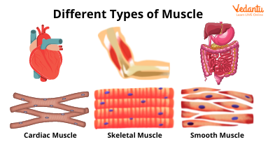
Different Types of Muscle
Skeletal Muscle
These are the most abundant muscles.
It is composed of muscle bundles (fascicles), kept intact by collagenous connective tissue called fascia.
Each muscle bundle contains a number of muscle fibres.
Each muscle fibre is lined by the plasma membrane called sarcolemma enclosing the sarcoplasm.
Muscle fibre is a syncytium as the sarcoplasm contains many nuclei.
Sarcoplasmic reticulum which is the endoplasmic reticulum of the muscle fibres is the storehouse of calcium ions.
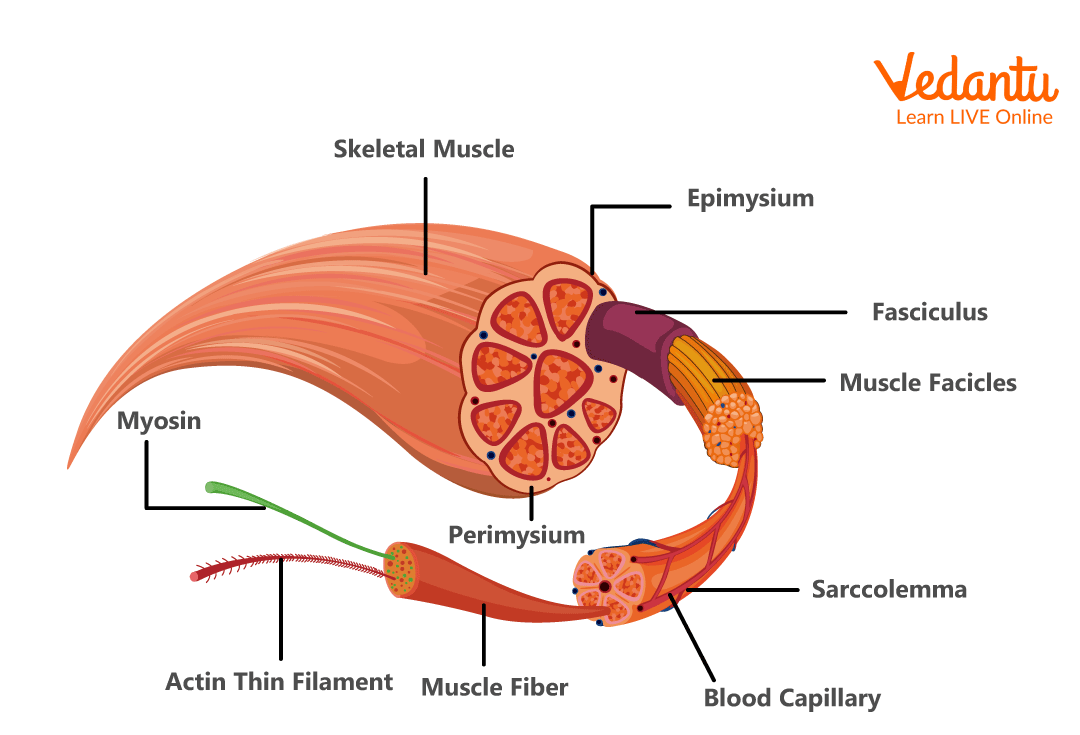
Diagram of Skeletal Muscle
Myofibrils are parallelly arranged by a large number of filaments in the sarcoplasm, which is the characteristic feature of the muscle fibre.
Alternate light and dark colour bands are present in each myofibril. These alternate bands gave the striated appearance to the myofibril.
There are two sorts of myofilaments present in a myofibril. Thin and thick filaments.
Thin filaments and Thick filaments need to join one another for muscle contraction.
Light bands contain actin and are called I-band (isotropic band) and dark bands contain myosin, called A-band (anisotropic band). Both the bands are available parallel with one another in a longitudinal style.
One myosin is surrounded by six actins, while one actin is surrounded by three myosins.
In the centre of every I-band is the elastic fibre called the 'Z' line.
In the A-band is a thin fibrous 'M' line.
The part of myofibrils between two progressive 'Z' lines is the functional unit of contraction called a sarcomere.
At the resting stage, thin filament overlaps thick filament. The part of the thick filament which is not overlapped by the thin filament is known as the ‘H-Zone’.
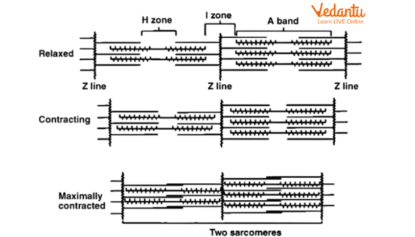
Relaxation and Contraction carried out by Sarcomere
Muscle Movement
The mechanism of muscle contraction is explained by the sliding mechanism hypothesis in which thin filaments slide over thick filaments.
Contraction of muscle starts with the signal sent by CNS through the motor neuron.
Neuron signals release neurotransmitters (Acetylcholine) to produce an action potential in the sarcolemma.
This causes the release of Ca2+ from the sarcoplasmic reticulum.
Ca2+ initiates actin which binds to the myosin head to form a cross bridge.
These cross bridges pull the actin fibres making them slide over the myosin fibres and consequently causing contraction.
Ca2+ is then gotten back to the sarcoplasmic reticulum which inactivates the actin. Cross bridges are broken and the muscles relax.
In this way, muscular movement takes place.
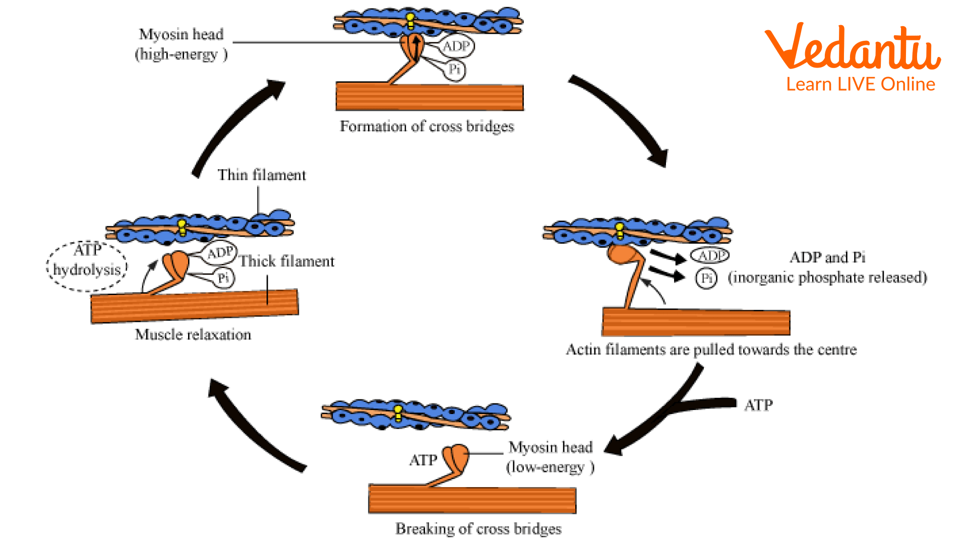
Mechanism of Muscle Contraction
Skeletal Movement
Structural framework of our body is provided by the skeletal muscle and brings about movement and locomotion.
Skeletal Muscle provides protection to the internal organs.
Bones and cartilage make up the skeletal system.
Tendons and ligaments are two forms of connective tissues that are also considered part of the system. Tendons connect bones to muscles, whereas ligaments connect bones to one other.
Bones:
In an adult human being, there are 206 bones in all.
Because of the calcium salts in the matrix, bones are hard, but cartilage includes chondroitin salts.
The human skeleton is divided into two sections: the axial skeleton, which has 80 bones, and the appendicular skeleton, which has 126 bones.
The skull, rib cage, and spinal column are all part of the axial skeleton, which itself is situated around the middle of the body.
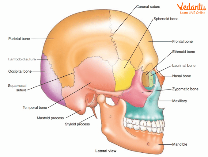
Axial Skeleton
The appendicular skeleton is made up of bones found in appendages such as the arms, legs, fingers, and toes.
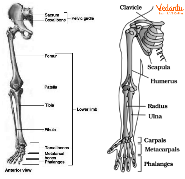
Appendicular skeleton
Joints
Joints are found in between the bones, also between the bone and cartilage.
Joints aid in locomotion and movement.
There are three different kinds of joints:
(i) Fibrous Joints:
They are immobile joints which don’t allow any movement.
They can be seen in the cranium.
(ii) Cartilaginous Joints:
They have limited movement, such as the intervertebral disc between two vertebrae in the spine.
Bones are held together by cartilage.
(iii) Synovial Joints:
Synovial Joints are movable joints.
They feature a fluid-filled synovial space between two bones that allows for a lot of movement.
Following are the types of synovial joints:
Pivot Joint (Present between Atlas and Axis)
Ball and Socket Joint (Shoulder)
Hinge Joint (Knee and Elbow)
Gliding Joint (Carpal)
Saddle Joint (in thumb, between carpal and metacarpal)
Conclusion
This article gives insight into the movement and locomotion skeletal system. It talks about the difference between locomotion and movement, the structure of the muscle, and the functional unit of the muscle which brings about the contraction and the relaxation of the muscle, thick and thin filaments and their composition, skeletal movement and muscular movement. It also covers different joints as well which work together in a coordinated manner to bring the locomotion and movement. It is a very important chapter of Biology.
FAQs on Movement and Locomotion Skeletal System for NEET
1. How many questions are asked from Locomotion and Movement in the examination?
This chapter in itself is a very important chapter of human physiology and covers essential topics and carries a strong weightage from the exam point of view. A student is advised to go through each diagram of the NCERT thoroughly and practise a lot of the labelling of the diagrams because diagrams surely play an essential role in the examination. Students must revise the diagrams and their labelling and should retain the concept in a tabular manner to avoid further confusion in the examination.
2. What are the important topics from Locomotion and Movement?
This is one of the most interesting and scoring chapters of human physiology. It has several important topics including different types of muscles, their properties and their location, through understanding the functional unit of muscle and its different components. There are many different confusing terms in the chapter to which students must pay attention. Mechanism of contraction and relaxation of muscle, different types of joints, axial and appendicular skeleton, and the total number of bones and muscles in the body are some of the important topics and students must revise them again and again.


