Revision Notes on Structural Organisation in Animals NEET 2025 - Free PDF Download
Structural Organisation in Animals is an important topic of the CBSE Class 11 Biology subject as well as NEET Biology syllabus. This chapter concentrates on topics such as surface animal tissues, organ and organ systems, structural organisation of frogs, structural organisation of earthworm, and structural organisation of cockroaches. It also discusses how the human body is made up of billions of cells that conduct a variety of jobs.
These Structural Organisation in Animals notes will help aspirants understand and remember the chapter better. The specialists have simplified the scientific ideas of this chapter. As a result, it will aid in your preparation for the NEET exams.
Note: 👉Get a Head Start on Your Medical Career with the NEET Rank and College Predictor 2025.
Access NEET Revision Notes Biology Structural Organisation in Animals
 Introduction

The cells of multicellular organisms are organised into tissues to carry out various functions.
Tissue: A group of cells and intercellular substances that act together to conduct a specific function.
Organ: A group of similar and dissimilar tissues in a living thing that has been organised and adapted to perform a specific function, including the heart, lung, kidney, or stomach.
An organ system is a combination of organs that work together to accomplish a single function. Each serves a specific purpose in the body but comprises distinct tissues.
All complex animals are composed of four basic tissue types.
Types of Animal Tissues
Epithelium Tissue:
They are closely packed cells with too little intercellular matrix, with one surface associated with exposure to air or internal fluid.
It lines the internal organs and cavities, covers the body's surface, acts as a barrier against mechanical damage, pathogens, and dehydration; and it creates a surface for molecule uptake, excretion, and transport.
There are two types of epithelium:
Simple epithelium
Stratified epithelium.
Simple Epithelium:
End-to-end arrangement of cells in a single layer. It is most commonly used to line body cavities, ducts, and tubes.
The simple epithelium's structure is divided into three types: squamous, cuboidal, and columnar.
Squamous Epithelium:
A thin layer of flattened cells with improper boundaries that would seem polygonal in surface view.
Because of their compact design, which resembles floor tiles, they are also identified as pavement epithelium.
It is responsible for the delicate lining of cavities like the mouth, oesophagus, nose, pericardium, and alveoli, as well as the lining of blood vessels.
They participate in activities like the development of a diffusion barrier and the creation of the tongue and skin covering.
To avoid wear and tear, the squamous epithelium is organised in many layers in the skin.
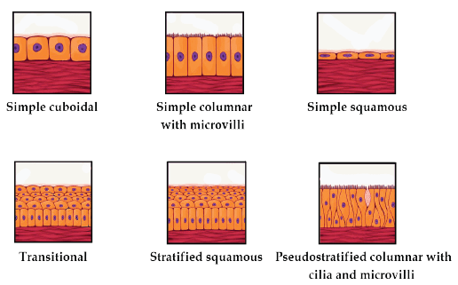
Types of Epithelial Tissues
Cuboidal Epithelium:
A coat of cube-like cells that emerge in sections as squares but are hexagonal upon that free surface.
It is present in thyroid vesicles, kidney tubules, and glands.
It is in charge of forming the germinal epithelium inside the gonads.
It helps with absorption, secretion, excretion, and also mechanical support.
Microvilli are found in the proximal convoluted tubule epithelium of a kidney's nephron. Microvilli are finger-like extensions on the surface of epithelial cells. They contribute to the absorption process.
Columnar Epithelium:
A thin layer of tall, thin cells in which the nuclei can be found at the base of the cell near the basement membrane.
On the free surface, microvilli may be present.
They are present in the stomach and intestine linings and aid in absorption and secretion.
Ciliated epithelium:
The cuboidal or columnar epithelium has fine, hair-like protoplasmic extensions on the surface.
It can be found in the kidneys, trachea, as well as fallopian tubes.
Cilia direct the movement of particles or mucus across the epithelium.
Glandular Epithelium:
Cells that have been genetically modified to secrete.
Columnar or cuboidal shapes are possible.
There are single, isolated glandular cells that are equivalent to goblet cells in the alimentary canal as well as a multicellular group of glandular cells, just like in the salivary gland.
Exocrine glands produce substances into the body via ducts or tubes. These glands secrete saliva, mucus, milk, oil, earwax, digestive enzymes, and other cell products.
They lack ducts and are endocrine (ductless glands).
Hormones are their byproducts and are effectively released into the fluid surrounding the gland.
1. Stratified or Compound Epithelium:
These cells are multi-layered and have more than one layer of cells.
They play a diminished role in secretion and absorption.
They are primarily secure against mechanical and chemical stress.
They are found on the skin's dry surface, the moist surface of the mouth cavity, the inner membrane of salivary gland ducts, the pharynx, and the pancreatic ducts.
Cellular Junctions:
Junctions are specialised structures that structurally and functionally connect individual cells.
Tight, adhering, and gap junctions are the three types.
Tight junctions are specialised structures that keep substances from seeping across the tissue.
Adhering junctions are a type of specialised structure that help to connect adjacent cells with each other.
Gap Junctions are specialised structures that enable cells to interact with one another. They connect neighbouring cells' cytoplasms, enabling the rapid transport of ions, and small and large molecules.
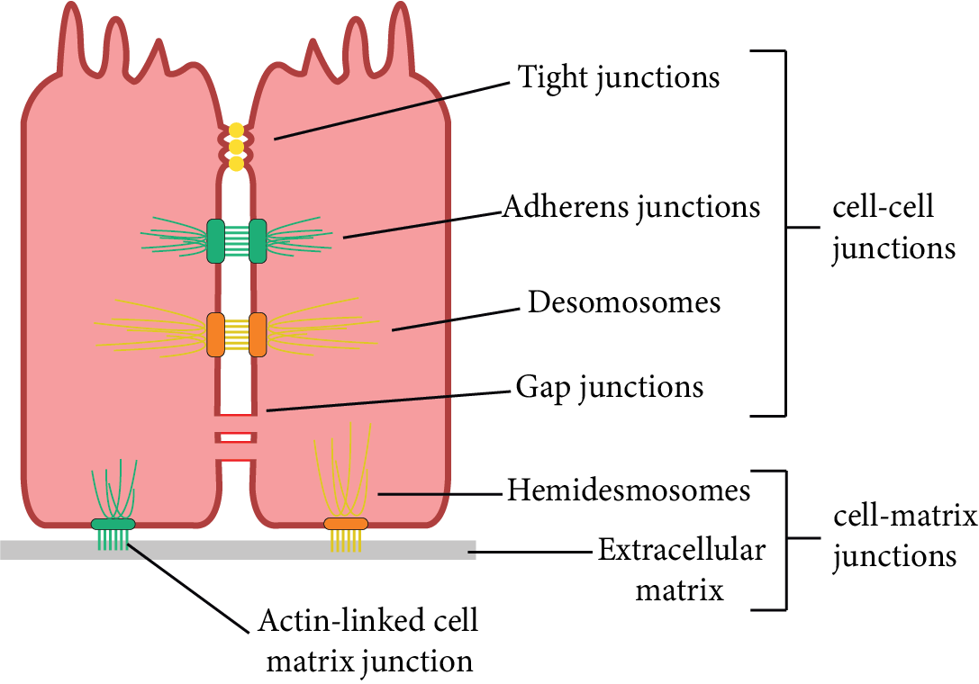
Types of Cell Junctions
Connective Tissue
The connective tissues of various types are found in the bodies of complex life forms. They are the most widely distributed.
They get their name because they play a unique function in the linking and assisting of body tissues/ organs.
They emerge in a variety of shapes and sizes, from soft to specialised.
Connective tissue is composed of widely spaced cells that are engrained in a matrix.
The nature of the matrix governs the function of the tissue.
Except for blood, cells in all connective tissues naturally produce structural proteins named collagen or elastin that provide the tissue with strength, elasticity, and flexibility.
The matrix or ground substance is created as part of the cell's secretion of modified polysaccharides.
The three major types of connective tissue are:
Loose connective tissue
Dense connective tissue
Specialised connective tissue.
Loose Connective Tissue:
Areolar, adipose, and reticular tissues are semi-fluid ground substances with loosely arranged cells and fibres.
Areolar Tissue:
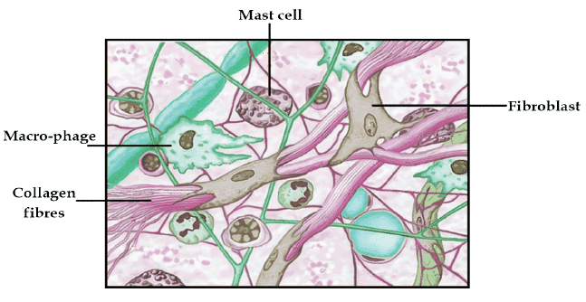
Areolar Tissue
This category of connective tissue is the most common in the body.
This tissue can be present inside the organs, between organs and skin, in nerves, bone marrow, and around blood vessels.
It is regularly used as an epithelial support framework.
Fibroblasts, macrophages, and mast cells comprise it.
Fibroblasts produce and secrete two types of fibres: elastic yellow fibres and inelastic white fibres.
It interconnects tissues in an organ.
It aids in the replacement of damaged body tissues.
Allergies are a concern with mast cells.
Adipose Tissue:
This tissue can be found mostly beneath the skin.
The cells are built to hold fat.
It is composed of adipocytes, which seem to be oval and round cells containing fat globules.
It prevents heat loss from the body by acting as insulation.
It aids in the backup of excess energy such as fats.
It acts as a cushioned shock absorber around vital organs including kidneys, eyeballs, heart, etc.
Reticular Tissue:
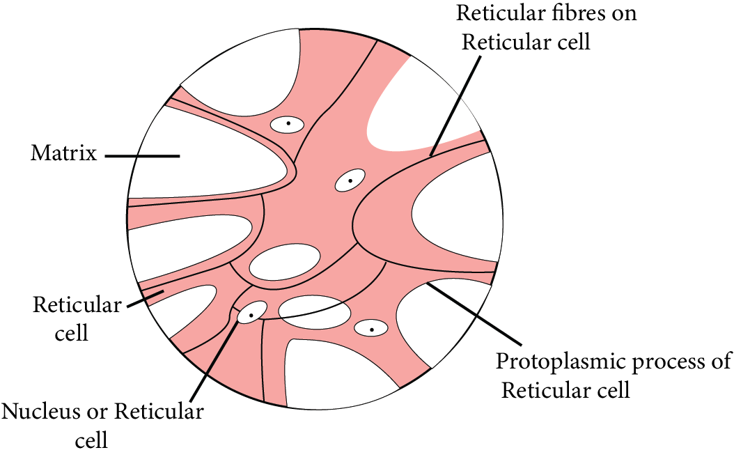
Reticular Tissue
The cells of this tissue are star-shaped and comprise a network-like architecture.
Reticulin protein makes up the fibres.
The spleen, tonsils, lymph nodes, and other organs contain it.
It is in charge of the development of lymphoid tissues in the body.
Reticular tissue inside the bone marrow aids in the formation of blood cells.
Dense Connective Tissue:
Cells, fibres, and fibroblasts are packed together with a little matrix.
Tissue orientation can be either regular or irregular, resulting in the terms dense regular as well as dense irregular tissues.
Dense Regular Connective Tissues:
Collagen fibres are found between many parallel fibre bundles.
There are rows of collagen fibres visible.
Tendons and ligaments are a couple of examples.
Tendons are fibrous white tissues. The fibroblasts are positioned almost continuously. It's stiff and unyielding.
A ligament attaches a skeletal muscle to a bone. Ligaments are made of yellow elastic tissue. The fibroblasts are all over the place. It's a tough but malleable tissue. It links one bone to the next.
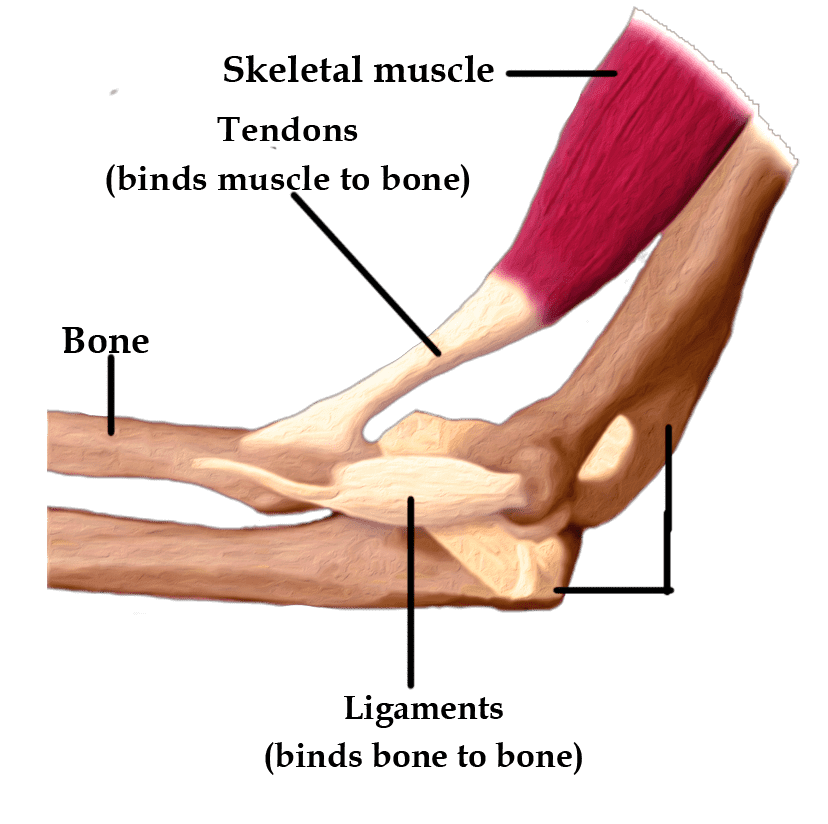
Tendon and Ligament
Dense irregular connective tissues:
In this case, the fibroblasts and fibres are directed differently.
The skin is made up of these dense irregular connective tissues.
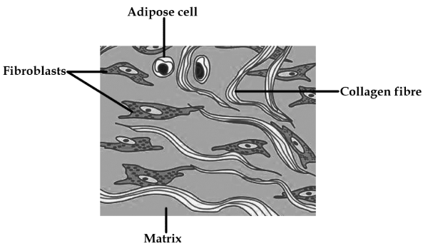
Dense Irregular Tissue
Specialised Connective Tissue:
Connective tissue which has been prepared to carry out a specific function is referred to as specialising connective tissue.
It is made up of bone, cartilage, and lymph.
Bone:
A category of skeletal tissue is bone.
It provides structural support to the body.
It is made of a hard, non-pliable mixture of calcium phosphate, calcium carbonate, and ossein protein.
Collagen protein is also a major component of bone.
The matrix is made up of layers identified as lamellae. The lamellae seem like crescentic rings around Haversian canals, which are longitudinal canals.
Lacunae are fluid-filled spaces between ring-shaped lamellae.
Bone cells, also defined as osteocytes, are found in the lacunae.
Bones provide support and protection for soft tissues and organs.
The weight of the body is supported by limb bones, like the long bones of the legs. To produce movement, they also communicate with the skeletal muscles attached to them.
Blood cells are produced in the bone marrow of some bones.
Cartilage:
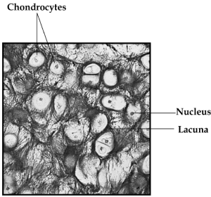
Cartilage Tissue
Cartilage is also a form of skeletal tissue.
This tissue is tough and elastic, and it's not as hard as bone.
The matrix is both solid and pliable, with the ability to withstand compression.
Cartilage cells, also defined as chondrocytes, are secreted in small holes within the matrix.
The elasticity is due to the presence of the protein chondrin.
Cells are extensively separated, and fibres reinforce the matrix.
It is in charge of vertebrate embryonic skeletons. Bones eventually replace the major portion of the cartilage.
Cartilage can be found in places like the tip of the nose, adult limbs, and hands, outer ear joints, etc. It can also be noticed between adjacent vertebral column bones.
Blood:
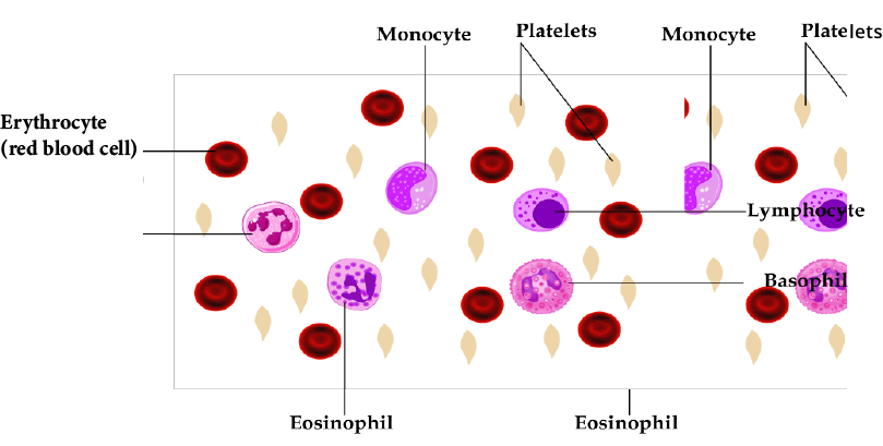
Blood Tissue
A fluid connective tissue composed of plasma and blood cells that represent the primary circulating fluid but also aids in the long-distance travel of various substances.
It's the most tender connective tissue there is.
Plasma is a liquid component that contains nearly 90% water.
Proteins, hormones, salts, and other materials used to carry digested food, gases, excretory products, and so on make up the remaining 10%.
Blood cells include red blood cells (RBCs), white blood cells (WBCs), and platelets.
Blood has a protein called albumin which is present in its serum in abundant amounts.
Blood's primary function is to transport oxygen and nutrients to the lungs and tissues, to produce blood clots to prevent excessive blood loss, to transfer cells and antibodies that fight infection, and to transport waste material to the kidneys and liver.
Muscular Tissue
Muscular tissue is distinguished by its tendency to contract and relax, enabling it to perform physical work.
Muscles are composed of numerous long cylindrical fibres organised in parallel arrays.
Myofibrils, which are fine fibrils, make up the fibres.
When muscle fibres are stimulated, they contract. They then relax and come back to a normal, uncontracted state in unison.
Their action makes the body or parts of the body shift in order to adapt to changes in the environment and retain the various body parts' positions.
Skeletal, smooth, and cardiac muscle cells are the three types of muscle cells.
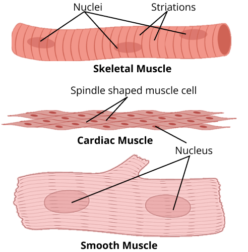
Skeletal, Smooth, and Cardiac muscles
Skeletal Muscles:
It is inextricably linked to the skeletal bones.
They are striated and arranged in a parallel pattern.
A tough connective tissue sheath surrounds several bundles of muscle fibres.
They are also identified as voluntary muscles since they are controlled by the mind.
Cells are unbranched and also have numerous nuclei.
Smooth Muscles:
Cells in smooth muscle taper at both free ends to frame a spindle or fusiform shape.
They're not striped.
They are held together by cell junctions, which are wrapped in a connective tissue sheath.
Nuclei do not exist in cells.
Because their activity is not controlled by consciousness, they are also identified as involuntary muscles.
Smooth muscle is located in the walls of visceral organs like the intestines, uterus, and stomach. These muscles can also be located in the lining of passageways, such as the arteries and veins of the cardiovascular system, as well as urinary, respiratory, and reproductive tracts. In addition, smooth muscle can be found in the eyes, where it changes the size of the iris as well as the shape of the lens.
Cardiac Muscles:
The cardiac muscle is a kind of contractile muscle tissue that can only be found in the heart.
Cell junctions are structures that link up the plasma membranes of cardiac muscle cells, allowing them to stick together. These are basically communication intersections.
Intercalated discs at certain fusion points allow cells to contract as a unit. When one cell gets a signal, the cells surrounding it are also stimulated to contract.
They are muscles that are involuntary.
They are uninucleate and branched.
These muscles contract and relax in a rhythmic pattern throughout life.
Nervous Tissue
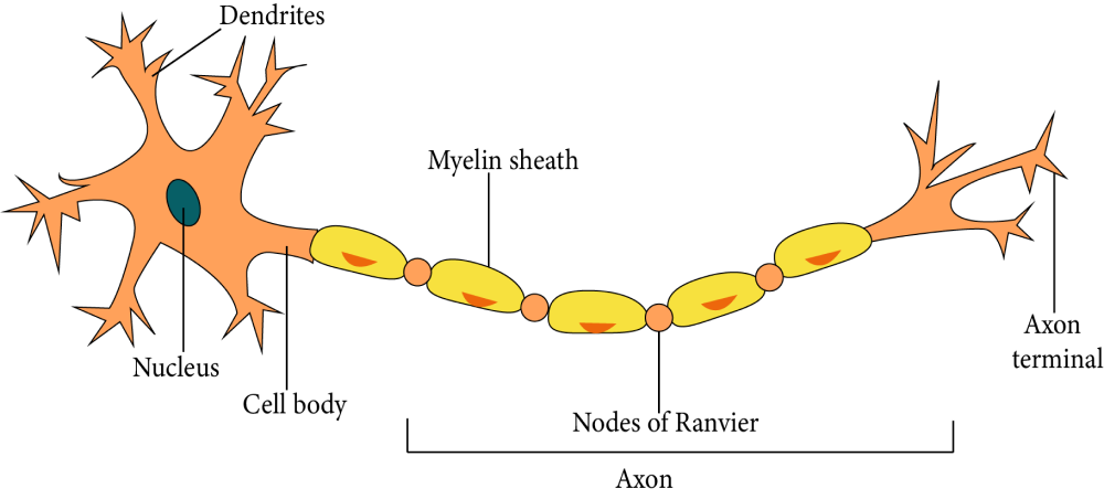
Nerve Cell
Neural tissue is composed of densely populated cells known as neurons and is specialised for nerve impulse conduction.
Neurons are found all over the brain, spinal cord, and nerves.
Neuroglial cells protect and nourish neurons.
Neuroglia makes up more than half of all neural tissue in our bodies.
The neuron is composed of three parts: the cyton, dendrites, and axon.
Cyton: The cell body that contains a nucleus in the centre and cytoplasm with differentiated deeply stained granules known as Nissl's granules.
Dendrites: These are cytoplasmic processes that are short and branched. They take impulses from receptors as well as other neurons and transmit them to the cyton.
Axon: A single long process that transmits impulses from one neuron to the next.
Nerve fibres can be myelinated or unmyelinated.
When a nerve cell is stimulated, an electrical impulse is generated that travels speedily along the plasma membrane. When this impulse achieves the ends of the neuron, it triggers events that either stimulate or inhibit neighbouring neurons.
Organ and the Organ System
The basic tissue types of the body are arranged in various ways to construct organs.
A group of these organs will then connect with one another to establish organ systems in multicellular organisms.
The body's organisation into tissues, organs, and organ systems is required for the body to function more efficiently.
It also facilitates the improved coordination of the actions of an organism's millions of cells.
Every body part in our body is composed of at least one type of tissue. Our heart, for example, is composed of all four tissue types: epithelial, connective, muscular, and neural.
Earthworm
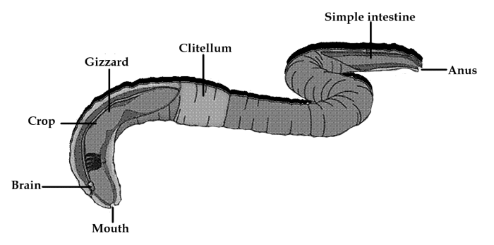
Earthworm
An earthworm is a reddish-brown terrestrial invertebrate that chooses to live in the top layer of moist sand.
Worm casting is a faecal deposit that can be used to identify them.
Morphology:
An earthworm's body is long and cylindrical. More than a hundred short and identical segments called metamers make up the body.
A dark median mid-dorsal line represents the dorsal blood vessel on its body's dorsal surface.
The existence of genital openings distinguishes the ventral surface (pores).
The mouth and the prostomium are at the front end. The prostomium is a groove that covers the mouth.
The prostomium has a sensory role and is used as a wedge to push open soil cracks so that the earthworm can crawl through them.
The mouth is located in the peristomium (buccal cavity), the first segment of the body.
The segments 14-16 of a mature worm are enclosed by a prominent dark patch of glandular tissue named the clitellum.
There are three sections to the body: preclitellar, clitellar, and postclitellar.
On the ventrolateral edges of the intersegmental grooves, there seem to be four pairs of spermathecal apertures. That is, they appear between the fifth and ninth segments.
A single female genital opening can be found in the mid-ventral line of the 14th segment.
On the ventrolateral edges of the 18th segment, there are two male genital pores.
On the body's surface, multiple small pores are defined as nephridiopores open.
Nephridiopores: Many life forms, including flatworms and annelids, have excretory organs called nephridiopores.
Rows of S-shaped setae cover each body segment. Setae are absent at the first, last, and clitellum segments of the body.
They are found in the epidermal pits in the centre of each segment. Setae already have the ability to expand and contract. Their primary function is to move around.
Anatomy:
A thin non-cellular cuticle protects the earthworm's body wall from the outside, preceded by the epidermis, two muscle layers, and an inner coelomic epithelium.
The Digestive System:
The alimentary canal refers to a straight duct that runs from the top of the body to the bottom.
A mouth at the tube's end unlocks into the buccal cavity (1-3 segments).
The muscular pharynx comes next.
There is a minor oesophagus between the fifth and seventh segments.
It develops into a muscular gizzard (8-9 segments).
Between segments 9 and 14, the stomach is situated.
The earthworm feeds on decaying leaves as well as organic matter mixed with soil.
The stomach is home to calciferous glands. They work to neutralise humic acid, which is found in humus.
The intestine begins with the 15th segment and finishes with the last segment. A pair of short, conical intestinal caeca bulging from the intestine on the 26th segment.
A typhlosole is present which is an internal median fold of the dorsal wall and extends from the 26th to the last segment except the last 23rd-25th segments. It is a unique characteristic of the intestine. This increases the effective absorption area of the intestine.
The undigested food moves to the rectum, where water is absorbed as well as the undigested food is ejected through the anus as castings. The anus, a small rounded aperture, connects the alimentary canal to the surroundings.
The ingested soil is rich in organic matter. It passes through the gastrointestinal tract.
Digestive enzymes in the digestive tract degrade organic food into smaller absorbable units. These smaller molecules are assimilated and utilised via intestinal membranes.
The Circulatory System:
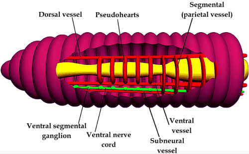
Circulatory System of Earthworm
Earthworm has a closed blood vascular system made up of blood vessels, capillaries, and the heart.
It shows a closed circulatory system, suggesting that blood is restricted to the heart and blood vessels.
Contractions restrict blood flow in one direction.
Smaller blood vessels transport nutrients and oxygen to the gut, nerve cord, and body wall.
The blood, blood vessels, capillaries, and the heart comprise the circulatory system. Because of the contractions, blood flows in only one direction.
The blood glands that produce blood cells and haemoglobin, are located in the fourth, fifth, and sixth segments. Blood cells lack haemoglobin and are phagocytic. Haemoglobin, in dissolved form, is found in blood plasma.
There are three major blood vessels that carry blood to the earthworm's organs:
The aortic arches work similarly to the human heart. Five pairs of aortic arches are responsible for pumping blood into the dorsal and ventral blood vessels.
The dorsal blood vessels are in charge of transporting blood to the earthworm's head.
The ventral blood vessels are in charge of transporting blood to the back.
The Respiratory System:
Earthworms lack specialised breathing equipment.
Respiratory exchange occurs via the moist body surface. Through the moist skin, gases are swapped directly into the bloodstream.
The Excretory System:
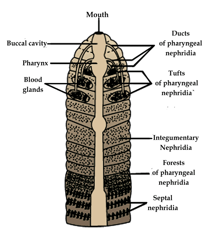
Excretory System of Earthworm
Earthworm excretory organs are referred to as nephridia.
They are coiled tubules that are segmentally arranged.
The three different types of nephridia are septal, integumentary, and pharyngeal nephridia.
Septal nephridia are found at the final intersegmental septa on segment 15. They are present on both sides. They allow for intestine access.
Integumentary nephridia are connected to the covering of the body wall from segment 3 to the last segment that reaches the body surface.
Pharyngeal nephridia emerge as three paired small clusters in the 4th, 5th, and 6th segments.
The structure of such various types of nephridia is nearly identical.
The volume and proportions of bodily fluids are regulated and modulated by nephridia.
A nephridium begins as a funnel. It drains excess fluid from the coelomic chamber. The funnel is linked to a tubular component. Waste is transported into the digestive tube by this.
The Nervous System:
It is composed of ganglia arranged in segments along the ventral paired nerve cord. In the anterior region, the nerve cord splits (3rd and 4th segments). After that, it folds around the pharynx.
Finally, it forms a nerve ring by connecting the dorsal cerebral ganglia.
Sensory input is integrated by the cerebral ganglia and the other nerves in the nerve ring. They are also in charge of the body's muscular reactions.
Organs responsive to light and touch are part of the sensory system. They exist in the form of receptor cells. They can tell the difference between light levels and aid in the interpretation of ground vibrations. There aren't any eyes.
In the anterior portion of the worm, there are specialised chemoreceptors (taste receptors) that respond to chemical stimuli.
The Reproductive System:
The earthworm is hermaphrodite (bisexual), which also means that it has both the testicles and the ovaries.
There are two pairs of testes in the tenth and eleventh segments.
Their vasa deferentia enlarges all the way to the 18th segment, at which they join the prostatic duct.
There are two types of accessory glands. They appear in the 17th and 19th segments.
The vasa deferentia is the main prostate and spermatic duct. It communicates with the outside world through a pair of male genital pores. These openings can be observed on the 18th segment's ventrolateral side.
There are four spermatheca pairs that can be identified in segments 6–9. Each segment includes one spermatheca pair. They obtain and store spermatozoa during copulation.
Spermatheca: A female or hermaphrodite animal's sac or receptacle. This sac holds the male's sperm until the eggs are prepared to get fertilised.
The inter-segmental septum between the 12th and 13th segments contains a pair of ovaries.
Ovarian funnels are placed beneath the ovaries and open into the ovaries. The oviduct is connected to the ovarian funnels. In the 14th segment, they later merge and open on the ventral surface to create a single median female genital pore.
During mating, two worms exchange sperm with one another.
One worm comes across another. When the worms' reproductive holes are opposite each other, mating occurs. These gonadal openings are known as spermatophores. They are used to send and receive sperm packets.
Clitellum glands produce cocoons. These cocoons contain mature sperm, egg cells, and also nutritive fluid.
Fertilisation and ova progression occurs within the cocoons.
The cocoons are then released into the soil. The fertilised ova, along with the worm's cocoon, then falls off. The cocoon is just where worm embryos grow.
After about three weeks, each cocoon creates two to twenty baby worms. Four are created on average.
Earthworms develop without the development of larvae.
Economical Importance:
Earthworms are also identified as "farmer's friends" because they dig holes in the soil, which helps make it porous. This enables growing plant roots to invade and breathe.
Vermicomposting is accomplished by using earthworms. In this method, earthworms are used to decompose the organic and soil, boosting soil fertility.
They are used as fishing bait.
Cockroach
They are brown or black-bodied creatures of the Phylum Arthropoda's Class Insecta.
In tropical areas, brightly colored cockroaches of red, yellow, and green have been observed.
They measure 0.-7-6 cm in length.
They have tall antennae, legs, and an upper body wall outgrowth that covers up their head.
They are nocturnal omnivores who prefer damp conditions.
They infiltrate homes and serve as disease vectors.
Morphology:
Periplaneta americana is the most predominant species of cockroach, measuring 33-54 mm in length.
Males' wings are seen to enlarge beyond the abdomen.
Their bodies are divided into three sections: the head, thorax, and abdomen.
An exoskeleton protects the entire body. It is hard and well-made of chitin.
Sclerites are hardened exoskeleton plates that link each segment via a thin and pliable arthrodial membrane.
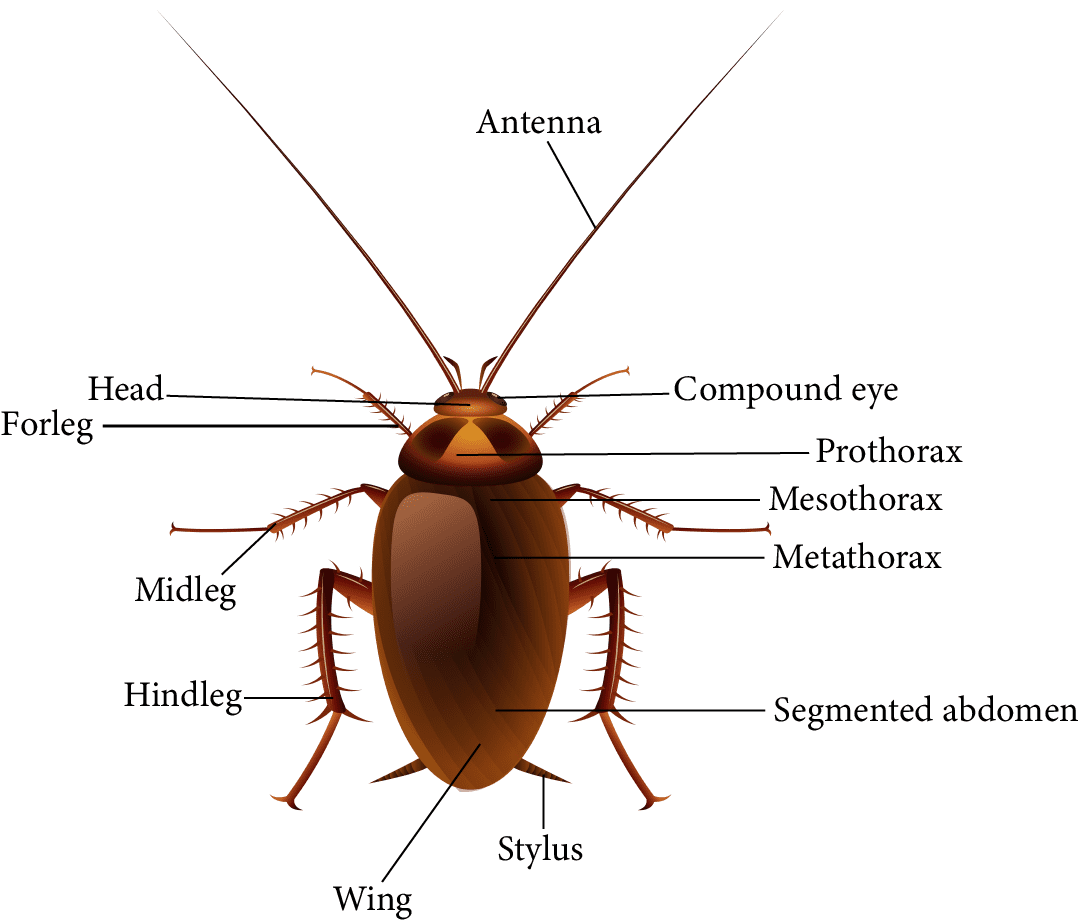
Morphology of Cockroach
Head:
The triangular head is created by merging six segments and found anterior at perpendicularly to the longitudinal body axis.
He has amazing mobility in all directions, thanks to a flexible neck.
On the head capsule, there are two compound eyes.
Antennae grow from membranous sockets located in front of the eyes.
Sensory receptors in antennas sense environmental changes.
The anterior end of the head's mouth parts has specialised protrusions for biting and chewing.
A labrum (upper lip), a labium (lower lip), two mandibles, and two maxillae make up the mouthparts.
A median flexible lobe provides the tongue (hypopharynx) inside the cavity encapsulated by the mouthparts.
Thorax:
There are three sections to the thorax: the prothorax, the mesothorax, and the metathorax.
The neck is the structure that attaches the head to the thorax.
The neck is a prothoracic extension that is short.
There are two walking legs in each thoracic segment.
Two pairs of wings are present.
The first pair is formed by the mesothorax, whereas the second is formed by the metathorax.
Tegmina: The forewings or mesothoracic wings of a cockroach. They have a dark, thick, and leathery appearance. They help hide the hind wings when at rest.
Metathoracic wings are the hind wings. They are transparent and membranous and are used to fly.
Abdomen:
The abdomen is split into ten sections.
The seventh sternum in females is shaped like a boat.
The brood or genital pouch is formed by the 7th, 8th, and 9th sterna.
The female gonopore, spermathecal pores, as well as collateral glands are located in the anterior part of the brood.
A genital pouch is located at the back of the abdomen in males. The 9th and 10th terga bind the genital pouch dorsally. The ventral genital pouch is connected by the 9th sternum.
The anus is found in the dorsal region of the brain. The ventral region houses the male genital pore. The pouch also contains the gonapophysis.
Males have two anal styles that are short and threadlike. They do not exist in females.
Anal cerci: A pair of hinged filamentous structures located on the 10th segment of both males and females.
Anatomy:
Alimentary Canal/ Digestive System Anatomy:
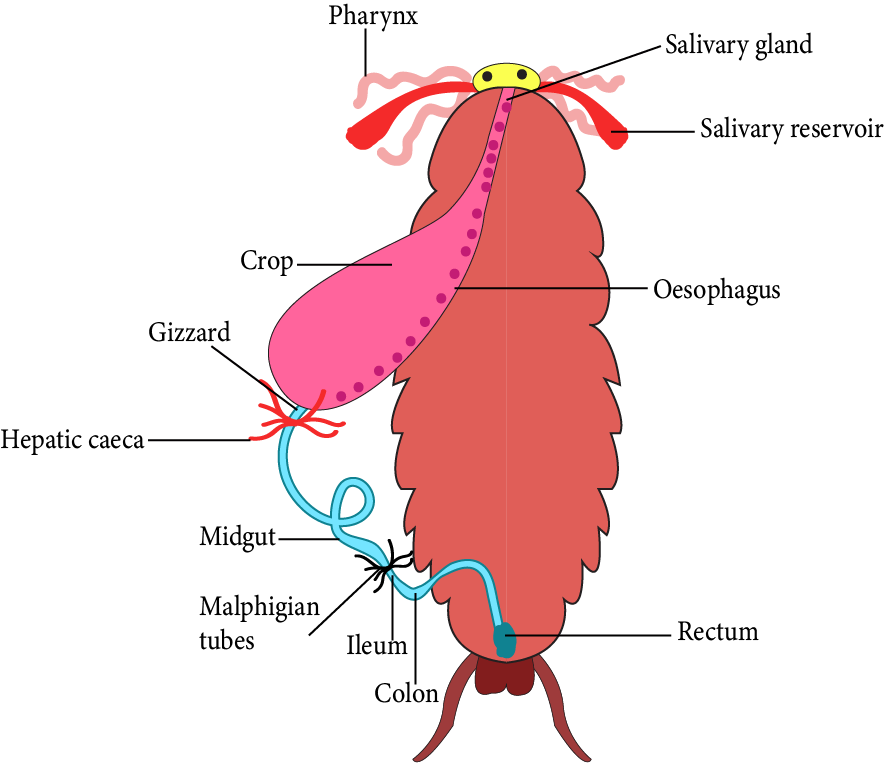
Digestive System of Cockroach
The alimentary canal is categorised into three parts: the foregut, midgut, and hindgut.
It is divided into the following sections:
Mouth: It terminates in a short tubular pharynx.
The pharynx joins the narrow tubular oesophagus.
A crop is a sac-like structure located at the oesophageal end. It's used to store food.
The crop is followed by the gizzard or proventriculus. It has a thick outer layer of circular muscles and a thick inner layer of cuticle that shapes six highly tough outer plates known as teeth. The gizzard helps to grind food particles.
The cuticle runs the length of the foregut.
The midgut is composed of 6-8 blind tubules called hepatic or gastric caeca. It can be found at the foregut-midgut junction. It secretes digestive juice.
Malpighian tubules are a ring of 100-150 yellow-colored fine filamentous tubules that are noticed at the midgut-hindgut junction. They help with the removal of excretory materials from the hemolymph.
Hindgut: It is bigger than the stomach. The ileum, colon, and rectum are the three sections. The anus provides access to the rectum.
Blood Vascular or Circulatory System:
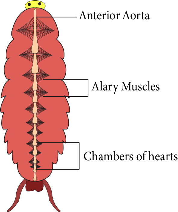
Circulatory System of Cockroach
Hemolymph: A fluid that spreads within the body of an arthropod while staying in physical touch with the animal's tissues, equivalent to blood in vertebrates. It is composed of fluid plasma in which hemocytes, or hemolymph cells, are suspended.
The cockroach's circulatory system is open.
The hemocoel has a collection of small blood vessels that reach the central body cavity. Visceral organs are found in the hemocoel. They are encircled by and in direct contact with the hemolymph.
The hemolymph is colourless. It consists of plasma and haemoglobin. Both plasma and haemoglobin are colourless.
The heart is an enlarged muscular tube that extends along the thoracic and abdominal mid-dorsal lines. It consists of 13 funnel-shaped chambers. Ostia can be observed on both sides of the heart.
The sinuses send blood to the heart via the Ostia. It is then pumped anteriorly again to the sinuses.
Ostia: A pair of small holes at the back of each heart chamber. It helps blood to flow into the heart.
Respiratory System:
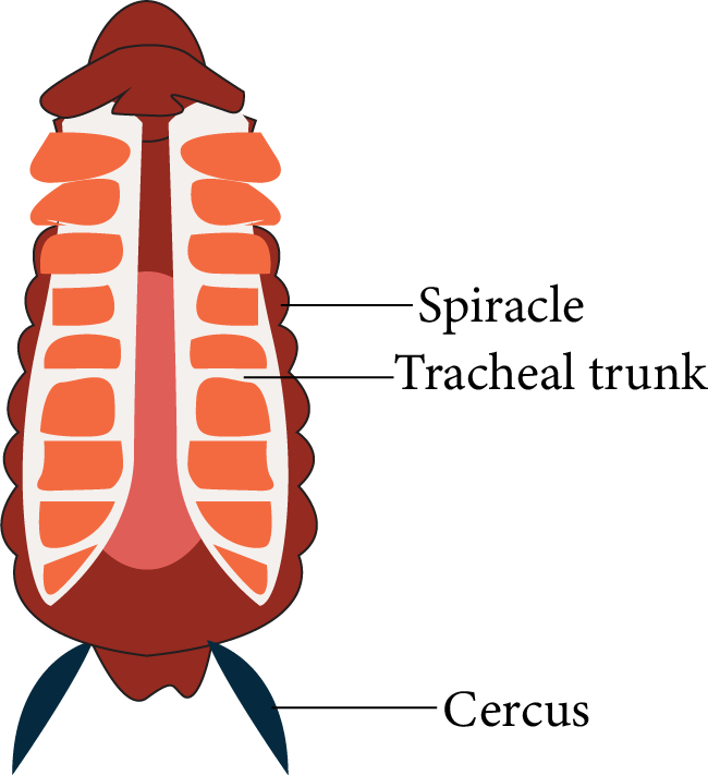
Respiratory system of cockroach
It consists of a cluster of tubes equivalent to the trachea that end in ten pairs of small openings known as spiracles. These spiracles can be noticed side to side in their bodies.
Spiracles are external respiratory system openings located in certain animal species. Insects, spiders, some fishes, and whale species contain them.
Spiracles help to transport oxygen to the internal respiratory parts. Internal respiratory organs vary between animals, such as whale lungs and insect tracheae.
Tracheoles are branching tubes in the trachea that transfer oxygen from the atmosphere to all areas of the body. The method by which gases transfer is known as diffusion.
Sphincters: Muscle-like cells that regulate mouth opening.
Excretory System:
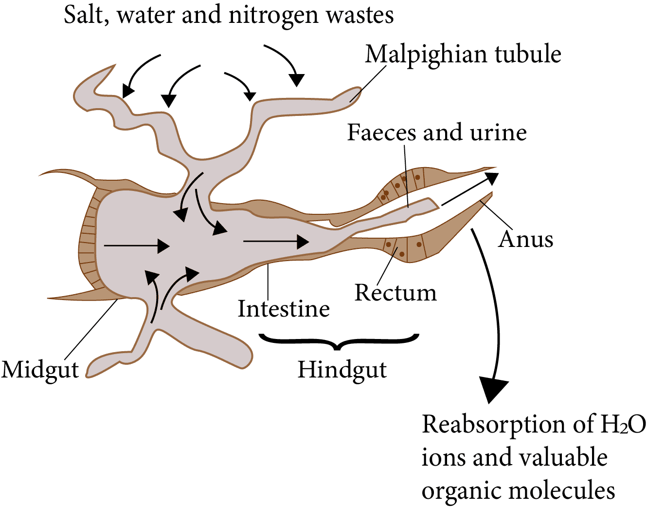
Excretory System of Cockroach
Malpighian tubules, which are layered by glandular and ciliated cells help to excrete.
They digest nitrogenous waste and transform it into uric acid, which is removed from the body through the hindgut. Thus, this insect is referred to as uricotelic.
The excretion of the Malpighian tubules is aided by the fat body, nephrocytes, and urecose glands.
Nervous system:
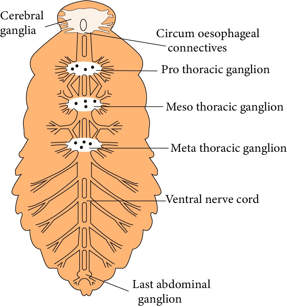
The nervous system is composed of ganglia. These ganglia are fused and segmentally organised. They are organised on the ventral side by paired vertical connectives.
There are three ganglia in the thorax. Six ganglia are found in the abdomen.
A cockroach's nervous system is dispersed all across the body, with the nervous system separated between the head as well as the ventral region.
As a result, a cockroach can survive without its head for up to a week.
The supra-oesophageal ganglion, which delivers nerves to the antennae and compound eyes, represents the brain in the head region.
The supraesophageal ganglion is also referred to as the "arthropod brain". It is the primary component of an arthropod's central nervous system, especially in insects.
Antennae, maxillary palps, eyes, labial palps, anal cerci, and other sense organs are included.

Nervous System of Cockroach
Compound eyes are identified on the head's dorsal surface. Each eye is made up of about 2000 hexagonal ommatidia.
These ommatidia help a cockroach receive various pictures of an object. This is known as mosaic vision.
It has increased sensitivity but decreased resolution. It is especially sensitive at night. As a result, it is known as nocturnal vision.
Ommatidia are arthropod compound eyes found in insects, crustaceans, as well as millipedes. Each ommatidium consists of a group of photoreceptor cells encircled by support and pigment cells. A transparent cornea protects the ommatidium's outer surface.
Reproductive System:
Cockroaches reproduce in two sexes.
Both sexes' reproductive organs are fully developed.

Male and Female Reproductive System of Cockroach
Male Reproductive System: It consists of two testes, one on each lateral side between the fourth and sixth abdominal segments.
Each testis produces Vas deferens.
The seminal vesicles allow Vas deferens to enter the ejaculatory duct.
The male gonopore is accessed via the ejaculatory duct.
The gonopore is the entrance of the ejaculatory duct in males. It is on the underside of the anus.
Auxiliary reproductive gland: In the sixth and seventh abdominal segments, there is a mushroom-shaped gland.
Male gonapophysis, also known as phallomeres, are the external genitalia.
The male gonopore is surrounded by a chitinous irregular structure called a phallomere.
The sperms are bundled together and deposited in the seminal vesicles. These bundles are known as spermatophores. During copulation, they are expelled.
Spermatophores are clusters of sperm that adhere to one another and are expelled during ejaculation.
Female reproductive System: It is composed of two large ovaries that are located laterally in the second to 6th abdominal segment.
Each ovary contains eight ovarian tubules. These ovarian tubules are known as ovarioles. Each ovariole is composed of a series of developing ovaries.
The oviducts of each ovary connect to create a single median oviduct. It is also known as the vagina. It provides entry to the genital cavity. A pair of spermatheca allows access to the genital chamber. It's in the sixth segment.
Spermatheca: a sperm-storing sac in the female reproductive tract. It is found in a wide range of lower animals, most notably insects.
Spermatophores are the units that transport sperm.
Fertilised eggs are housed in capsules called ootheca.
The ootheca is a big dark reddish to the blackish-brown chamber about 8mm in length.
They are either set down or attached to an appropriate surface. The surface should be near a source of food and have a high moisture content.
Females produce 9-10 ootheca on average. Every ootheca has between 14 and 16 eggs.
The development of eggs is a paurometabolous process.
A nymph stage is involved in paurometabolous development, which is a type of partial metamorphosis.
Nymph: A stage of imperfect metamorphosis in which the immature form resembles the adult but is smaller. Moulting allows the nymph to mature into an adult. Wing pads can be found in the nymph stage, just prior to the adult stage, however, only adults have wings.
Tips to Remember:
Cells, tissues, organs and organ systems split up the work in a way that ensures the survival of the body as a whole and exhibit division of labour.
A tissue is defined as a group of cells along with intercellular substances performing one or more functions in the body.
Epithelia are sheet-like tissues lining the body's surface and its cavities, ducts and tubes.
Epithelia have one free surface facing a body fluid or the outside environment. Their cells are structurally and functionally connected at junctions.
Diverse types of connective tissues bind together, support, strengthen, protect, and insulate other tissue in the body.
Soft connective tissues consist of protein fibres and various cells arranged in a ground substance.
Cartilage, bone, blood, and adipose tissue are specialised connective tissues.
An earthworm is a reddish-brown terrestrial invertebrate that chooses to live in the top layer of moist sand.
An earthworm's body is long and cylindrical. More than a hundred short and identical segments called metamers make up the body.
A thin non-cellular cuticle protects the earthworm's body wall from the outside, preceded by the epidermis, two muscle layers, and an inner coelomic epithelium.
Cockroaches are brown or black-bodied creatures of the Phylum Arthropoda's Class Insecta.
In tropical areas, brightly colored cockroaches of red, yellow, and green have been observed.
Periplaneta americana is the most predominant cockroach species, measuring 33-54 mm in length.
The alimentary canal of cockroaches is categorised into the foregut, midgut, and hindgut.
Importance of Structural Organisation in Animals Class 11 Notes for NEET
Students understandably might get frightened or nervous about the exam, especially if it's NEET. And due to that nervousness, they might find it challenging to concentrate on the exams. However, with these Structural Organisation in Animals Class 11 notes for NEET, students can now prepare better for the exam.
The notes simplify every aspect of this chapter. You would get an explanation of how animals are made up of several cell kinds. The structural organisation of various organisms varies. Tissues are formed when distinct cells unite. Examine how these notes can help you enhance your understanding of this chapter. Make your study and revision sessions more rewarding by using these notes.
Also, learn how tissue is a collection of comparable cells that work together with intercellular chemicals to execute a specific function in multicellular beings. You would get a simplified explanation stating how these tissues are combined to form organs. Learn more about this organisation by reading the notes on Structural Organisation in Animals.
On reading the Structural Organisation in Animals notes, you will be able to thoroughly understand the concepts of this chapter and answer questions easily. By using these notes, you may create a solid conceptual basis.
Benefits of Structural Organisation in Animals Class 11 Notes PDF Download
If you are still wondering why you should download the PDF, check out these reasons:
You would get to practice question answers that will let you understand the pattern in which NEET question papers are set. Learning this will automatically calm your nervousness about the upcoming competitive exam.
Vedantu's subject matter specialists have discussed in detail all essential topics and subtopics concerning the structural organisation of animals in this chapter for your ease of understanding. Every concept in this explanation will be separately taught so that you may answer the related question during the exams and score higher.
Understand the meaning and significance of animal tissues in depth. Use Vedantu's revision notes for NEET Structural Organisation in Animals Class 11 to easily grasp the structural organisation in-depth and other terminologies.
Prepare for the NEET test by answering questions about these themes. Follow the instructions in the Structural Organisation in Animals Class 11 notes for NEET to learn how Biology in every animal works.
Download Structural Organisation in Animals Notes PDF
Complete your preparation for appearing in NEET by downloading the PDF version of the revision notes for this chapter. Discover how animal tissues are classified as nervous, epithelial, muscular, or connective tissues. Learn how the question pattern would be and how to answer the questions accurately. The Structural Organisation in Animals Class 11 notes for NEET will also help ease your nervousness about the examination so that you can completely focus on ranking the scoreboard.
NEET Biology Revision Notes -Chapter Pages
NEET Biology Chapter-wise Revision Notes | |
Structural Organisation in Animals Notes | |
Other Important Links
Other Important Links for NEET Structural Organisation in Animals |
FAQs on Structural Organisation in Animals NEET Notes 2025
1. What are the different cell junctions found in tissues?
There are three different junctions in tissues
- Tight junctions,
- Gap junctions, and
- Adherens junctions.
2. What is an 'amphibian'?
Amphibians are an animal breed that can live on land as well as in water.
3. How are frogs beneficial for mankind?
Frogs consume insects and defend crops. They are a crucial link in the ecosystem's food web and food chains. Their legs can also be eaten.
4. What is adipose tissue?
Adipose tissue is also known as fat tissue. They are a type of connective tissue made up of adipocytes found mainly beneath the skin. Its primary role is to insulate the body and store energy as fat.


