


a)Draw the ray diagram showing the formation of an image by a compound microscope.
b) Show that in order to achieve large magnification in a compound microscope the magnitude of the focal length of objective and the eyepiece should be small.
Answer
587.1k+ views
Hint: To draw the ray diagram of the compound microscope, it basically consists of two convex lenses namely the objective that is kept near the specimen and the eyepiece that is kept at our least distance of distinct vision. While drawing the ray diagram it is to be considered the image formed by the objective lies within the focal length of the eyepiece and the focal length of the eyepiece is greater than that of the objective. Further will obtain an expression for magnification power and verify whether to increase magnification the focal length of the either lenses should be reduced.
Complete step by step answer:
The ray diagram of a compound microscope is shown below in Fig 1.
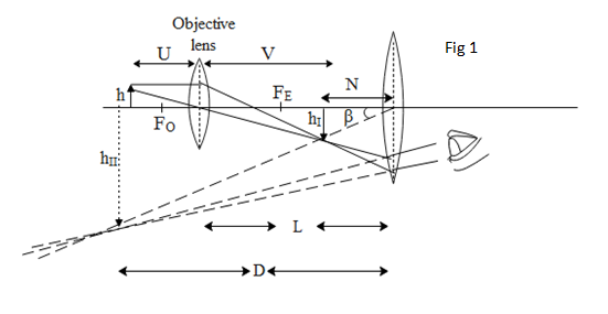

The magnifying power of the lens is defined as the ratio of the angle subtended by the image at the eye i.e. $\text{ }\!\!\beta\!\!\text{ }$ (as seen in Fig 1)to the angle subtended by the object at the eyepiece i.e. $\text{ }\!\!\alpha\!\!\text{ }$. (as seen in Fig 2) Mathematically written as,
The angular magnification of the image is given by, $m=\dfrac{\begin{align}
& \text{Angle Subtended by the image at least } \\
& \text{at the eyepiece} \\
\end{align}}{\begin{align}
& \text{Angle Subtended by the object at least } \\
& \text{at the eyepiece} \\
\end{align}}$
For small angle $\tan \theta \simeq \theta $ Hence, we can write the above equation as $m=\dfrac{\tan \beta }{\tan \alpha }$
From trigonometric definition of $\tan \theta $ we can write the above equation as,
$m=\dfrac{\dfrac{\text{height of the image }}{\text{-N}}}{\dfrac{\text{height of the object}}{-D}}=\dfrac{{{\text{h}}_{\text{I}}}}{\text{N}}\times \dfrac{\text{D}}{\text{h}}={{M}_{O}}{{M}_{E}}....(1)$
Where ${{M}_{O}}$is the magnification produced by the objective and ${{M}_{E}}$is the magnification produced by the eye piece.
${{M}_{O}}=\dfrac{\text{V}}{\text{U}}=\text{-}\dfrac{\text{L}}{{{\text{f}}_{\text{O}}}}\text{where L is the tube length and }{{\text{f}}_{\text{O}}}\text{ is the focal length of the objective}\text{.}$
An eyepiece acts as an simple microscope Hence magnification produced by the eyepiece is given by,
${{\text{M}}_{\text{E}}}\text{=}\dfrac{\text{D}}{{{\text{f}}_{\text{E}}}}\text{+1}$ . Substituting the magnifications of both the objective and the eyepiece in equation 1 we get,
$\begin{align}
& m={{M}_{O}}{{M}_{E}} \\
& m=\text{-}\dfrac{\text{L}}{{{\text{f}}_{\text{O}}}}\left( \dfrac{\text{D}}{{{\text{f}}_{\text{E}}}}\text{+1} \right) \\
\end{align}$
If we see the above equation the magnification of the compound microscope can be increased if we decrease the focal length of both the objective as well as the eye piece as they vary inversely.
Note: In actual practice each of the objectives and the eyepiece consists of a combination of lenses. To eliminate chromatic aberration, an objective consists of two lenses in contact. To reduce spherical aberration and chromatic aberration an eye piece consists of two lenses separated by a certain distance.
Complete step by step answer:
The ray diagram of a compound microscope is shown below in Fig 1.
The magnifying power of the lens is defined as the ratio of the angle subtended by the image at the eye i.e. $\text{ }\!\!\beta\!\!\text{ }$ (as seen in Fig 1)to the angle subtended by the object at the eyepiece i.e. $\text{ }\!\!\alpha\!\!\text{ }$. (as seen in Fig 2) Mathematically written as,
The angular magnification of the image is given by, $m=\dfrac{\begin{align}
& \text{Angle Subtended by the image at least } \\
& \text{at the eyepiece} \\
\end{align}}{\begin{align}
& \text{Angle Subtended by the object at least } \\
& \text{at the eyepiece} \\
\end{align}}$
For small angle $\tan \theta \simeq \theta $ Hence, we can write the above equation as $m=\dfrac{\tan \beta }{\tan \alpha }$
From trigonometric definition of $\tan \theta $ we can write the above equation as,
$m=\dfrac{\dfrac{\text{height of the image }}{\text{-N}}}{\dfrac{\text{height of the object}}{-D}}=\dfrac{{{\text{h}}_{\text{I}}}}{\text{N}}\times \dfrac{\text{D}}{\text{h}}={{M}_{O}}{{M}_{E}}....(1)$
Where ${{M}_{O}}$is the magnification produced by the objective and ${{M}_{E}}$is the magnification produced by the eye piece.
${{M}_{O}}=\dfrac{\text{V}}{\text{U}}=\text{-}\dfrac{\text{L}}{{{\text{f}}_{\text{O}}}}\text{where L is the tube length and }{{\text{f}}_{\text{O}}}\text{ is the focal length of the objective}\text{.}$
An eyepiece acts as an simple microscope Hence magnification produced by the eyepiece is given by,
${{\text{M}}_{\text{E}}}\text{=}\dfrac{\text{D}}{{{\text{f}}_{\text{E}}}}\text{+1}$ . Substituting the magnifications of both the objective and the eyepiece in equation 1 we get,
$\begin{align}
& m={{M}_{O}}{{M}_{E}} \\
& m=\text{-}\dfrac{\text{L}}{{{\text{f}}_{\text{O}}}}\left( \dfrac{\text{D}}{{{\text{f}}_{\text{E}}}}\text{+1} \right) \\
\end{align}$
If we see the above equation the magnification of the compound microscope can be increased if we decrease the focal length of both the objective as well as the eye piece as they vary inversely.
Note: In actual practice each of the objectives and the eyepiece consists of a combination of lenses. To eliminate chromatic aberration, an objective consists of two lenses in contact. To reduce spherical aberration and chromatic aberration an eye piece consists of two lenses separated by a certain distance.
Recently Updated Pages
Master Class 12 Economics: Engaging Questions & Answers for Success
Master Class 12 Physics: Engaging Questions & Answers for Success
Master Class 12 English: Engaging Questions & Answers for Success
Master Class 12 Social Science: Engaging Questions & Answers for Success
Master Class 12 Maths: Engaging Questions & Answers for Success
Master Class 12 Business Studies: Engaging Questions & Answers for Success
Trending doubts
Which are the Top 10 Largest Countries of the World?
What are the major means of transport Explain each class 12 social science CBSE
Draw a labelled sketch of the human eye class 12 physics CBSE
Why cannot DNA pass through cell membranes class 12 biology CBSE
Differentiate between insitu conservation and exsitu class 12 biology CBSE
Draw a neat and well labeled diagram of TS of ovary class 12 biology CBSE