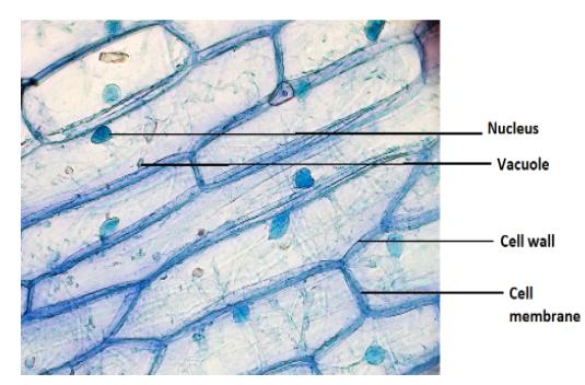
AIM: To prepare stained temporary mount of onion peel cells and to record observations and draw labelled diagrams.
MATERIALS REQUIRED: Onion, plain slides, coverslip, watch glass, needles, forceps, brush, blade, safranin, blotting paper, glycerine and compound microscope.
Answer
510k+ views
Hint: Staining is the method used extensively in the microscopy to make the mount/specimen visible under the microscope. By staining onion peel, it will make the cell wall more transparent in an onion cell. It adds contrast when seen through a microscope. Methylene Blue is a blue dye that can be used to colour blood, bacteria, acidic or protein-rich cell structures such as the nucleus, ribosomes, and endoplasmic reticulum.
Complete answer:
For preparing a temporary mount of the onion, the dye used is saffranine. Saffranine is extensively used in cytology. It will stain the cell nuclei red.
Procedure:
- Bend a slice of onion to extract the translucent membranous structure known as the onion epidermal peel. Remove the peel from its inner side using a forcep.
- In a watch glass, immerse the peel in water.
- To stain the watch glass holding the peel, add a few drops of stain safranin.
- Now, with the aid of a brush, wash the leaf peel and move it to a clean slide.
- Blotting paper may be used to remove excess water from the slide covering the peel.
- Add a drop of glycerine to this slide and position the coverslip in such a way that air bubbles do not enter.
- Blot away any excess glycerine with blotting paper.
- Examine the slide with a microscope.
Observation:
- It is possible to see a large number of rectangular cells with distinct cell walls.
- Cytoplasm is visible as a thin film of dark-coloured material on the inner surface of the cell wall.
- The cell contains a large central vacuole.
- Each cell has a deeply stained round body known as the nucleus.
Conclusion:As cell walls and large vacuoles are clearly observed in all the cells, the cells placed for observation are plant cells.

Note: We conclude
- Onion epidermal peel is made up of rectangular shaped cells. A nucleus, a central vacuole, a thin layer of cytoplasm, and a cell wall make up each cell.
- Since each cell has cell walls and a large prominent vacuole, the cells under observation are plant cells.
Complete answer:
For preparing a temporary mount of the onion, the dye used is saffranine. Saffranine is extensively used in cytology. It will stain the cell nuclei red.
Procedure:
- Bend a slice of onion to extract the translucent membranous structure known as the onion epidermal peel. Remove the peel from its inner side using a forcep.
- In a watch glass, immerse the peel in water.
- To stain the watch glass holding the peel, add a few drops of stain safranin.
- Now, with the aid of a brush, wash the leaf peel and move it to a clean slide.
- Blotting paper may be used to remove excess water from the slide covering the peel.
- Add a drop of glycerine to this slide and position the coverslip in such a way that air bubbles do not enter.
- Blot away any excess glycerine with blotting paper.
- Examine the slide with a microscope.
Observation:
- It is possible to see a large number of rectangular cells with distinct cell walls.
- Cytoplasm is visible as a thin film of dark-coloured material on the inner surface of the cell wall.
- The cell contains a large central vacuole.
- Each cell has a deeply stained round body known as the nucleus.
Conclusion:As cell walls and large vacuoles are clearly observed in all the cells, the cells placed for observation are plant cells.

Note: We conclude
- Onion epidermal peel is made up of rectangular shaped cells. A nucleus, a central vacuole, a thin layer of cytoplasm, and a cell wall make up each cell.
- Since each cell has cell walls and a large prominent vacuole, the cells under observation are plant cells.
Recently Updated Pages
Master Class 7 English: Engaging Questions & Answers for Success

Master Class 7 Maths: Engaging Questions & Answers for Success

Master Class 7 Science: Engaging Questions & Answers for Success

Class 7 Question and Answer - Your Ultimate Solutions Guide

Master Class 9 General Knowledge: Engaging Questions & Answers for Success

Master Class 9 Social Science: Engaging Questions & Answers for Success

Trending doubts
The value of 6 more than 7 is A 1 B 1 C 13 D 13 class 7 maths CBSE

Convert 200 Million dollars in rupees class 7 maths CBSE

List of coprime numbers from 1 to 100 class 7 maths CBSE

AIM To prepare stained temporary mount of onion peel class 7 biology CBSE

The plural of Chief is Chieves A True B False class 7 english CBSE

Write a letter to the editor of the national daily class 7 english CBSE


