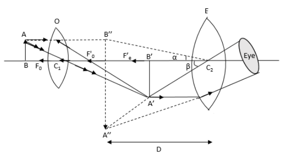
Draw a labelled ray diagram of an image formed by a compound microscope, when the final image lies at the least distance of distinct vision (D).
Answer
570k+ views
Hint:First recall the arrangement of objective and eyepiece lenses in the compound formation. Also, recall the process of image formation of an object in the compound microscope. For this process, draw the ray diagram of the compound microscope in which the image is formed at the least distance of distinct vision.
Complete step by step answer:
The ray diagram an image formed by a compound microscope, when the final image lies at the least distance of distinct vision (D) is as follows:

In the above ray diagram, O is the objective lens and E is the eyepiece lens.In the above ray diagram of the compound microscope, AB is the object, A’B’ is the image formed by the objective lens and A’’B’’ is the image formed by the eyepiece lens. The Focal length of the objective lens is ${F_o}$ and the focal length of the eyepiece lens is Fe. The distance of least distinct vision is shown by D.
Additional information:
The magnification of the image formed by the compound microscope for image formed at least distance of clear vision is given by
\[m = - \dfrac{L}{{{f_o}}}\left( {1 + \dfrac{D}{{{f_e}}}} \right) = {m_o}{m_e}\]
Here, \[L\] is the distance between the eyepiece and objective lens, \[{f_o}\] is the focal length of objective lens, \[{f_e}\] is the focal length of eyepiece, \[D\] is the least distance of clear vision, \[{m_o}\] is magnification of objective lens and \[{m_e}\] is magnification of eyepiece lens.
Note: The students should be careful while drawing the ray diagram because if the directions of the rays are not drawn properly, the final image formed by the eyepiece and objective lens in the ray diagram will not be in the proper position. Hence, the ray diagram drawn will not be correct.
Complete step by step answer:
The ray diagram an image formed by a compound microscope, when the final image lies at the least distance of distinct vision (D) is as follows:

In the above ray diagram, O is the objective lens and E is the eyepiece lens.In the above ray diagram of the compound microscope, AB is the object, A’B’ is the image formed by the objective lens and A’’B’’ is the image formed by the eyepiece lens. The Focal length of the objective lens is ${F_o}$ and the focal length of the eyepiece lens is Fe. The distance of least distinct vision is shown by D.
Additional information:
The magnification of the image formed by the compound microscope for image formed at least distance of clear vision is given by
\[m = - \dfrac{L}{{{f_o}}}\left( {1 + \dfrac{D}{{{f_e}}}} \right) = {m_o}{m_e}\]
Here, \[L\] is the distance between the eyepiece and objective lens, \[{f_o}\] is the focal length of objective lens, \[{f_e}\] is the focal length of eyepiece, \[D\] is the least distance of clear vision, \[{m_o}\] is magnification of objective lens and \[{m_e}\] is magnification of eyepiece lens.
Note: The students should be careful while drawing the ray diagram because if the directions of the rays are not drawn properly, the final image formed by the eyepiece and objective lens in the ray diagram will not be in the proper position. Hence, the ray diagram drawn will not be correct.
Recently Updated Pages
Master Class 12 Economics: Engaging Questions & Answers for Success

Master Class 12 Physics: Engaging Questions & Answers for Success

Master Class 12 English: Engaging Questions & Answers for Success

Master Class 12 Social Science: Engaging Questions & Answers for Success

Master Class 12 Maths: Engaging Questions & Answers for Success

Master Class 12 Business Studies: Engaging Questions & Answers for Success

Trending doubts
Which are the Top 10 Largest Countries of the World?

What are the major means of transport Explain each class 12 social science CBSE

Draw a labelled sketch of the human eye class 12 physics CBSE

Why cannot DNA pass through cell membranes class 12 biology CBSE

Differentiate between insitu conservation and exsitu class 12 biology CBSE

Draw a neat and well labeled diagram of TS of ovary class 12 biology CBSE


