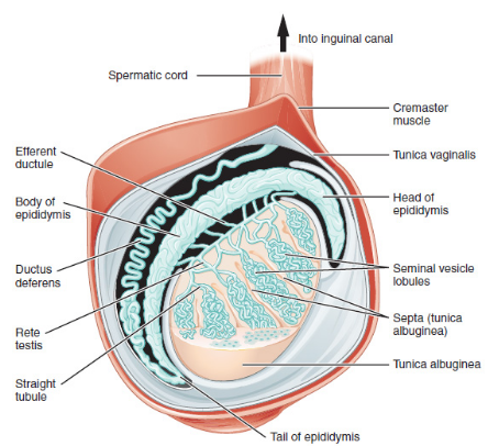
Explain with a neat labelled diagram the T.S. of mammalian testis.
Answer
578.1k+ views
Hint: In man, 1 pair testes are the main or primary reproductive organ. Size $4 - 5cm \times 2 - 3cm$. Both tests are located in small bags like structures situated below and outside the abdominal cavity known as scrotum or scrotal sac. The temperature of scrotum is 2-2.5$^ \circ C$ lesser than the body temperature. Each testis is attached to the wall of scrotal sac through flexible, elastic fibres. This group of fibre is called Gubernaculum or Mesorchium. Each testis is attached to the dorsal body wall of the abdominal cavity through a cord term as a spermatic cord.
Complete answer:

Internal structure of testis:-testis is covered by three Coats. outermost is tunica vaginalis. Middle coat is tunica albuginea and innermost tunica vasculosa. Tunica vaginalis-as a parietal and visceral layer. It covers the whole testis except its posterior border from where the testicular vessel and nerves enter the testis. Tunica albuginea-is a dense white fibrous coat covering the testis. All around The posterior border tunica albuginea is thickened to form vertical Septum called Mediastinum testis. Tunica vasculosa-is the innermost vascular coat of the testis line in testicular lobules.
Each lobule has 1 to 3 seminiferous tubules. Outer surface of the seminiferous tubule is composed of white fibrous connective tissue called tunica propria. while the inner surface is of cuboidal germinal epithelium. This epithelium is made up of spermatogenic cells which forms sperm by spermatogenesis. Some columnar cells are found in the layer of germinal epithelium called a sertoli cell. These provide nutrition to the germ cell, so they are also called sustentacular or nurse cells(these occur in mammals). Other functions of sertoli cells-
(1) They phagocytes the injured or dead sperm cells.
(2) They are bases of blood testis barriers.
(3) Sertoli cells produce inhibin and anti mullerian hormone.
(4) Sertoli cells can synthesize estrogen from testosterone.
Some endocrine cells are found between seminiferous tubules in intertubular space, these are called interstitial or Leydig cells. These cells secrete androgen that is testosterone. The testosterone from Leydig cells enter the seminiferous tubule by diffusion under the effect of ABP and promote spermatogenesis. Leydig's cells synthesize and secrete testicular hormones called androgen. Other immunologically competent cells are also present.
Note: Seminiferous tubules join together at the apices of the lobule to form straight tubules or tubuli recti which enter the mediastinum. Here they form a network of tubules called rete testis. Rete testis fuse to form 10 to 12 different efferent ductules called Vasa efferentia or ductuli efferentes. Total number of seminiferous tubules in each testis is about 750-1000.
Complete answer:

Internal structure of testis:-testis is covered by three Coats. outermost is tunica vaginalis. Middle coat is tunica albuginea and innermost tunica vasculosa. Tunica vaginalis-as a parietal and visceral layer. It covers the whole testis except its posterior border from where the testicular vessel and nerves enter the testis. Tunica albuginea-is a dense white fibrous coat covering the testis. All around The posterior border tunica albuginea is thickened to form vertical Septum called Mediastinum testis. Tunica vasculosa-is the innermost vascular coat of the testis line in testicular lobules.
Each lobule has 1 to 3 seminiferous tubules. Outer surface of the seminiferous tubule is composed of white fibrous connective tissue called tunica propria. while the inner surface is of cuboidal germinal epithelium. This epithelium is made up of spermatogenic cells which forms sperm by spermatogenesis. Some columnar cells are found in the layer of germinal epithelium called a sertoli cell. These provide nutrition to the germ cell, so they are also called sustentacular or nurse cells(these occur in mammals). Other functions of sertoli cells-
(1) They phagocytes the injured or dead sperm cells.
(2) They are bases of blood testis barriers.
(3) Sertoli cells produce inhibin and anti mullerian hormone.
(4) Sertoli cells can synthesize estrogen from testosterone.
Some endocrine cells are found between seminiferous tubules in intertubular space, these are called interstitial or Leydig cells. These cells secrete androgen that is testosterone. The testosterone from Leydig cells enter the seminiferous tubule by diffusion under the effect of ABP and promote spermatogenesis. Leydig's cells synthesize and secrete testicular hormones called androgen. Other immunologically competent cells are also present.
Note: Seminiferous tubules join together at the apices of the lobule to form straight tubules or tubuli recti which enter the mediastinum. Here they form a network of tubules called rete testis. Rete testis fuse to form 10 to 12 different efferent ductules called Vasa efferentia or ductuli efferentes. Total number of seminiferous tubules in each testis is about 750-1000.
Recently Updated Pages
Master Class 12 Economics: Engaging Questions & Answers for Success

Master Class 12 Physics: Engaging Questions & Answers for Success

Master Class 12 English: Engaging Questions & Answers for Success

Master Class 12 Social Science: Engaging Questions & Answers for Success

Master Class 12 Maths: Engaging Questions & Answers for Success

Master Class 12 Business Studies: Engaging Questions & Answers for Success

Trending doubts
Which are the Top 10 Largest Countries of the World?

What are the major means of transport Explain each class 12 social science CBSE

Draw a labelled sketch of the human eye class 12 physics CBSE

Why cannot DNA pass through cell membranes class 12 biology CBSE

Differentiate between insitu conservation and exsitu class 12 biology CBSE

Draw a neat and well labeled diagram of TS of ovary class 12 biology CBSE


