


What do the purkinje fibres connect to?
Answer
490.2k+ views
Hint: The rapid electric conduction in the ventricles is controlled by the His-Purkinje System (HPS). It relays electrical impulses from the atrioventricular node to muscle cells, coordinating ventricular contractions and ensuring proper cardiac pump function.
Complete answer:
In the case of humans, the size of the heart is approximately of a closed fist which is situated in between the lungs. The pumped blood carries all the nutrients and the oxygen to the whole body and all the metabolic waste like carbon dioxide is sent to the lungs.
In mammals and birds the heart is divided into four chambers: the upper part is known as atria which is further divided into right atria and left atria, and the bottom part is known as ventricle which is also further divided into right and left ventricle. Fishes have only two chambers while reptiles have three chambers. In heart valves are present which prevents the backflow of blood. Heart is covered with an outer protective layer which is known as pericardium. Heart wall has three layers: epicardium, myocardium and endocardium.
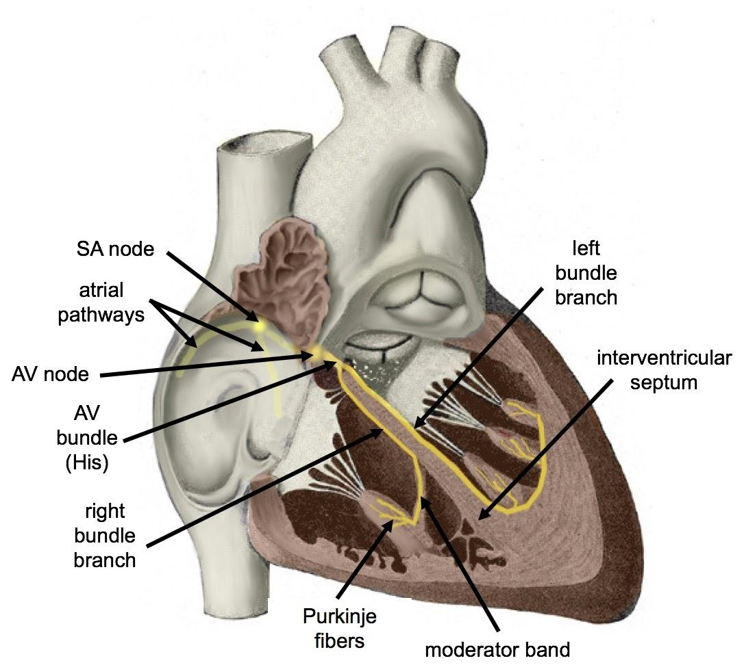
The heart pumps the blood with the help of a sinoatrial node known as SA node. It is also known as the pacemaker of the heart because it generates a current in the heart which causes the contraction in the walls of heart. This contraction causes the pumping of the heart through which blood flows to the whole body.
Purkinje fibres are the fibres which are present in the inner ventricular walls of the heart. They consist of electrically excitable cells which are specialized conducting fibres in nature. They have many mitochondria and few microfibrils and are larger than cardiomyocytes. These fibres allow the conduction system in the heart to maintain a consistent heart rhythm.
Purkinje fibers are a type of fiber found in the Purkinje system. Purkinje tissue (also known as subendocardial branches) is found in the heart's inner ventricular walls, just beneath the endocardium in a space known as the subendocardium. Purkinje fibers are electrically excitable cells that form specialized conducting fibers.
Purkinje fibres form an interweaving network on the endocardial surface of both ventricles by tying up with the ends of bundle branches. These fibres transmit the cardiac impulse to the entire left, right, and left ventricle endocardium almost simultaneously. The Purkinje fibres near the base of the ventricles have a tendency to be less intense.The inner third of the endocardium is only penetrated by the papillary muscle tips. When compared to normal myocardial fibres, it appears to be more resistant to ischemia.
Note:
If upstream conduction or pacemaking ability are compromised, Purkinje fibers can fire at a rate of 20-40 beats per minute. The SA node, on the other hand, can fire at 60-100 beats per minute in its normal state. Simply put, they produce action potentials at a slower rate than the sinoatrial node. Normally, this ability is disabled.
Complete answer:
In the case of humans, the size of the heart is approximately of a closed fist which is situated in between the lungs. The pumped blood carries all the nutrients and the oxygen to the whole body and all the metabolic waste like carbon dioxide is sent to the lungs.
In mammals and birds the heart is divided into four chambers: the upper part is known as atria which is further divided into right atria and left atria, and the bottom part is known as ventricle which is also further divided into right and left ventricle. Fishes have only two chambers while reptiles have three chambers. In heart valves are present which prevents the backflow of blood. Heart is covered with an outer protective layer which is known as pericardium. Heart wall has three layers: epicardium, myocardium and endocardium.
The heart pumps the blood with the help of a sinoatrial node known as SA node. It is also known as the pacemaker of the heart because it generates a current in the heart which causes the contraction in the walls of heart. This contraction causes the pumping of the heart through which blood flows to the whole body.
Purkinje fibres are the fibres which are present in the inner ventricular walls of the heart. They consist of electrically excitable cells which are specialized conducting fibres in nature. They have many mitochondria and few microfibrils and are larger than cardiomyocytes. These fibres allow the conduction system in the heart to maintain a consistent heart rhythm.
Purkinje fibers are a type of fiber found in the Purkinje system. Purkinje tissue (also known as subendocardial branches) is found in the heart's inner ventricular walls, just beneath the endocardium in a space known as the subendocardium. Purkinje fibers are electrically excitable cells that form specialized conducting fibers.
Purkinje fibres form an interweaving network on the endocardial surface of both ventricles by tying up with the ends of bundle branches. These fibres transmit the cardiac impulse to the entire left, right, and left ventricle endocardium almost simultaneously. The Purkinje fibres near the base of the ventricles have a tendency to be less intense.The inner third of the endocardium is only penetrated by the papillary muscle tips. When compared to normal myocardial fibres, it appears to be more resistant to ischemia.
Note:
If upstream conduction or pacemaking ability are compromised, Purkinje fibers can fire at a rate of 20-40 beats per minute. The SA node, on the other hand, can fire at 60-100 beats per minute in its normal state. Simply put, they produce action potentials at a slower rate than the sinoatrial node. Normally, this ability is disabled.
Recently Updated Pages
Master Class 11 Computer Science: Engaging Questions & Answers for Success
Master Class 11 Business Studies: Engaging Questions & Answers for Success
Master Class 11 Economics: Engaging Questions & Answers for Success
Master Class 11 English: Engaging Questions & Answers for Success
Master Class 11 Maths: Engaging Questions & Answers for Success
Master Class 11 Biology: Engaging Questions & Answers for Success
Trending doubts
One Metric ton is equal to kg A 10000 B 1000 C 100 class 11 physics CBSE
There are 720 permutations of the digits 1 2 3 4 5 class 11 maths CBSE
Discuss the various forms of bacteria class 11 biology CBSE
Draw a diagram of a plant cell and label at least eight class 11 biology CBSE
State the laws of reflection of light
Explain zero factorial class 11 maths CBSE