Write a note on eye defects and their correction.
Answer
521.1k+ views
Hint: The human eye is a sense organ which detects visible light and conveys this information to the brain. Eyes signal information which is used by the brain to obtain the perception of colour, shape, depth, movement, and other features. Defects in the eye leads to degradation of sight.
Complete answer:
There are many diseases, disorders and age-related changes that may affect the eyes and tamper with the vision. The most common refractive defects of vision are as given below:
1. Presbyopia
2. Myopia
3. Hypermetropia
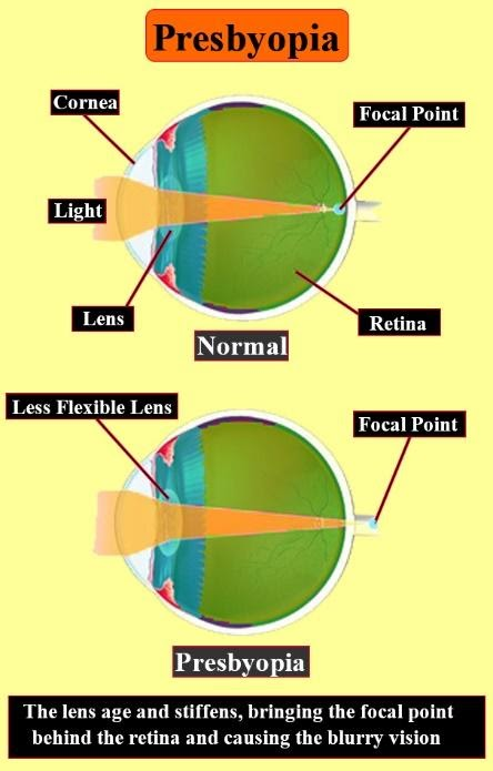
Presbyopia: With aging, the quality of vision worsens due to reduction in pupil size and the loss of accommodation or focusing capability. The area of the pupil commands the amount of light that can reach the retina. The extent of pupil dilation decreases with age and leads to a substantial decrease in the light received at the retina. Presbyopia occurs due to the gradual weakening of the ciliary muscles and the decreased flexibility of the eye lens.

Figure 1: Presbyopia
In order to overcome presbyopia, two main components of the visual system should be addressed: image capturing by the optical system of the eye and image processing in the brain.
Image capturing by the eye: Usage of corrective lenses, more specifically, convex lenses will increase the range of vision. Multifocal contact lenses can be used to correct vision for both the near and far-sightedness caused by presbyopia. Some people choose contact lenses for correcting one eye for near-sightedness and the other for far-sightedness with a method called monovision. Refractive surgery is another method to correct presbyopia. It is done to create multifocal corneas. PresbyLASIK is a type of multifocal corneal ablation LASIK procedure used to correct presbyopia. However, refractive surgery in some people decreases their visual acuity including postoperative glare, halos, ghost images and monocular diplopia.
Image processing by brain: A number of studies have claimed upgradation in near visual acuity by the use of training protocols based on perceptual learning and the eventual detection of low-contrast Gabor stimuli.
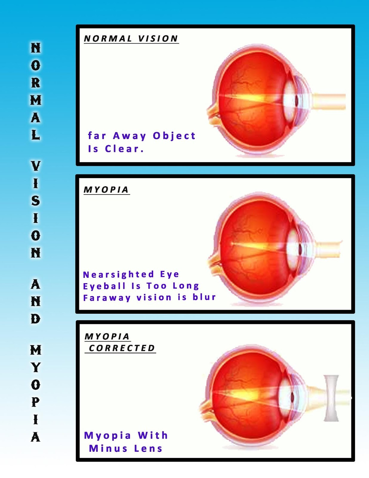
Myopia: Myopia, also called near-sightedness and short-sightedness is an eye defect where the light focuses in front of, instead of on, the retina. This causes the appearance of distant objects to be blurry while close objects to be normal. Headaches and eye strain are some consistent symptoms of myopia. Severe myopia is associated with an increased risk of retinal detachment, cataracts, and glaucoma. Causes of myopia include elongation of the eyeball and excessive curvature of the eye lens. Genetic linkage studies have identified 18 possible loci on 15 different chromosomes which are associated with myopia, but none of them are part of the genes that directly cause myopia. Instead of it being caused by a defect in a structural protein, defects in the control of these structural proteins might be the actual cause. A collaboration of all myopia studies worldwide identified 16 new loci for the refractive error in European ancestry, of which 8 were shared with Asians. The new loci include candidate genes with functions in neurotransmission, ion transport, retinoic acid metabolism and extracellular matrix remodelling along with eye development. The carriers of the high-risk genes are said to have a tenfold increased risk of myopia.

Figure 2: Myopia
Refractive surgery for myopia includes procedures which alter the corneal curvature of some structure of the eye or which add additional refractive means inside the eye. Photorefractive keratectomy, LASIK, Phakic intraocular lens, Orthokeratology, Intrastromal corneal ring segment are the refractive surgeries famous for the treatment of myopia.
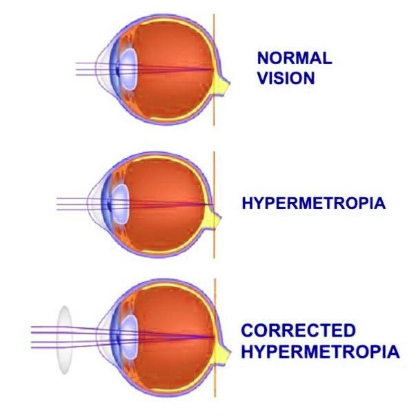
Hypermetropia: Far-sightedness, also termed as long-sightedness, hypermetropia, or hyperopia, is an eye defect where distant objects are seen clearly but near objects appear blurred. This blurred effect is due to incoming light being focused behind, instead of on, the retina wall caused by insufficient accommodation by the eye lens. It may occur when the axial length of the eyeball is short, or if the lens or cornea is flatter than normal.[2] Changes in the refractive index of the lens, alterations in its position or the total absence of lens also cause hypermetropia. Risk factors include a familial history of the condition, diabetes, certain medications and tumors around the eye. Functional hypermetropia occurs due to the paralysis of accommodation as evident in internal ophthalmoplegia, CN III palsy etc.
Eyeglasses used to correct hypermetropia or far-sightedness have convex lenses. Several surgeries can be administered to correct the condition. Laser procedures include Photorefractive keratectomy (removal of a minimal amount of the corneal surface), Laser assisted in situ keratomileusis (LASIK) (reshaping the cornea), Laser epithelial keratomileusis (LASEK) (uses alcohol to loosen the corneal surface), Epi-LASIK (uses epikeratome instead of alcohol), Laser thermal keratoplasty (LKR) (uses Thallium-Holmium-Chromium). IOL implantation and non-laser procedures (like Conductive keratoplasty, Automated lamellar keratoplasty, Keratophakia and epi-keratophakia).
Note:
In optics, the closest point at which an object can be brought into focus by the eye is called the eye's near point. A standard near point is a distance of 25 cm is typically assumed in the design of optical instruments. Lutein and zeaxanthin act as antioxidants that protect the retina and macula from oxidative damage due to high-energy light waves. Lack of lutein and zeaxanthin leads to macular degeneration or cataracts.

Figure 3: Hypermetropia
Complete answer:
There are many diseases, disorders and age-related changes that may affect the eyes and tamper with the vision. The most common refractive defects of vision are as given below:
1. Presbyopia
2. Myopia
3. Hypermetropia
Presbyopia: With aging, the quality of vision worsens due to reduction in pupil size and the loss of accommodation or focusing capability. The area of the pupil commands the amount of light that can reach the retina. The extent of pupil dilation decreases with age and leads to a substantial decrease in the light received at the retina. Presbyopia occurs due to the gradual weakening of the ciliary muscles and the decreased flexibility of the eye lens.

Figure 1: Presbyopia
In order to overcome presbyopia, two main components of the visual system should be addressed: image capturing by the optical system of the eye and image processing in the brain.
Image capturing by the eye: Usage of corrective lenses, more specifically, convex lenses will increase the range of vision. Multifocal contact lenses can be used to correct vision for both the near and far-sightedness caused by presbyopia. Some people choose contact lenses for correcting one eye for near-sightedness and the other for far-sightedness with a method called monovision. Refractive surgery is another method to correct presbyopia. It is done to create multifocal corneas. PresbyLASIK is a type of multifocal corneal ablation LASIK procedure used to correct presbyopia. However, refractive surgery in some people decreases their visual acuity including postoperative glare, halos, ghost images and monocular diplopia.
Image processing by brain: A number of studies have claimed upgradation in near visual acuity by the use of training protocols based on perceptual learning and the eventual detection of low-contrast Gabor stimuli.
Myopia: Myopia, also called near-sightedness and short-sightedness is an eye defect where the light focuses in front of, instead of on, the retina. This causes the appearance of distant objects to be blurry while close objects to be normal. Headaches and eye strain are some consistent symptoms of myopia. Severe myopia is associated with an increased risk of retinal detachment, cataracts, and glaucoma. Causes of myopia include elongation of the eyeball and excessive curvature of the eye lens. Genetic linkage studies have identified 18 possible loci on 15 different chromosomes which are associated with myopia, but none of them are part of the genes that directly cause myopia. Instead of it being caused by a defect in a structural protein, defects in the control of these structural proteins might be the actual cause. A collaboration of all myopia studies worldwide identified 16 new loci for the refractive error in European ancestry, of which 8 were shared with Asians. The new loci include candidate genes with functions in neurotransmission, ion transport, retinoic acid metabolism and extracellular matrix remodelling along with eye development. The carriers of the high-risk genes are said to have a tenfold increased risk of myopia.

Figure 2: Myopia
Refractive surgery for myopia includes procedures which alter the corneal curvature of some structure of the eye or which add additional refractive means inside the eye. Photorefractive keratectomy, LASIK, Phakic intraocular lens, Orthokeratology, Intrastromal corneal ring segment are the refractive surgeries famous for the treatment of myopia.
Hypermetropia: Far-sightedness, also termed as long-sightedness, hypermetropia, or hyperopia, is an eye defect where distant objects are seen clearly but near objects appear blurred. This blurred effect is due to incoming light being focused behind, instead of on, the retina wall caused by insufficient accommodation by the eye lens. It may occur when the axial length of the eyeball is short, or if the lens or cornea is flatter than normal.[2] Changes in the refractive index of the lens, alterations in its position or the total absence of lens also cause hypermetropia. Risk factors include a familial history of the condition, diabetes, certain medications and tumors around the eye. Functional hypermetropia occurs due to the paralysis of accommodation as evident in internal ophthalmoplegia, CN III palsy etc.
Eyeglasses used to correct hypermetropia or far-sightedness have convex lenses. Several surgeries can be administered to correct the condition. Laser procedures include Photorefractive keratectomy (removal of a minimal amount of the corneal surface), Laser assisted in situ keratomileusis (LASIK) (reshaping the cornea), Laser epithelial keratomileusis (LASEK) (uses alcohol to loosen the corneal surface), Epi-LASIK (uses epikeratome instead of alcohol), Laser thermal keratoplasty (LKR) (uses Thallium-Holmium-Chromium). IOL implantation and non-laser procedures (like Conductive keratoplasty, Automated lamellar keratoplasty, Keratophakia and epi-keratophakia).
Note:
In optics, the closest point at which an object can be brought into focus by the eye is called the eye's near point. A standard near point is a distance of 25 cm is typically assumed in the design of optical instruments. Lutein and zeaxanthin act as antioxidants that protect the retina and macula from oxidative damage due to high-energy light waves. Lack of lutein and zeaxanthin leads to macular degeneration or cataracts.

Figure 3: Hypermetropia
Recently Updated Pages
Master Class 11 Computer Science: Engaging Questions & Answers for Success

Master Class 11 Business Studies: Engaging Questions & Answers for Success

Master Class 11 Economics: Engaging Questions & Answers for Success

Master Class 11 English: Engaging Questions & Answers for Success

Master Class 11 Maths: Engaging Questions & Answers for Success

Master Class 11 Biology: Engaging Questions & Answers for Success

Trending doubts
One Metric ton is equal to kg A 10000 B 1000 C 100 class 11 physics CBSE

There are 720 permutations of the digits 1 2 3 4 5 class 11 maths CBSE

Discuss the various forms of bacteria class 11 biology CBSE

Draw a diagram of a plant cell and label at least eight class 11 biology CBSE

State the laws of reflection of light

Explain zero factorial class 11 maths CBSE


