


Explain the working of compound microscope by focal length of convex lens:
Answer
516.9k+ views
Hint: The above question requires knowledge of concepts of compound microscope and properties of convex lenses. The compound microscope uses two convex lenses with short focal lengths to produce magnified images of extremely small objects. These are very popular in the study of microorganisms.
Complete step-by-step solution:
A compound microscope is an optical instrument that uses two convex lenses with short focal lengths to observe extremely magnified images of small objects. The picture of a small object can be magnified up to 1000 times using a compound microscope.
When a tiny object is positioned just outside the focus of a compound microscope's objective lens, a simulated, inverted, and highly magnified image of the object is created at the shortest distance of distinct vision from the eye kept close to the eye piece.
Working:
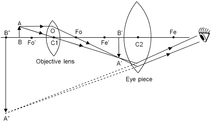
The figure depicts a ray diagram of a compound microscope in action. A small magnifying object AB is set in front of the objective lens just outside its primary focus ${F_o}'$. In this case, the compound microscope's objective lens O creates a true, inverted, and enlarged image of the object A'B'.
Now A'B' serves as an object for the eyepiece E, which has been changed to position A'B' between the optical center C2 and the eye piece's focus ${F_e}'$. The eyepiece now creates a simulated, reversed, and strongly magnified image A”B”. After adjusting the final image A”B” at the least distance of distinct vision of 25 cm from the eye, this final image A”B” is seen by our eye kept close to the eye.
Note:A magnifying glass is another name for a basic microscope. It's a convex lens with a short focal length that's used to see magnified views of tiny objects. A compound microscope is an optical instrument that uses two convex lenses with short focal lengths to observe extremely magnified images of small objects.
Complete step-by-step solution:
A compound microscope is an optical instrument that uses two convex lenses with short focal lengths to observe extremely magnified images of small objects. The picture of a small object can be magnified up to 1000 times using a compound microscope.
When a tiny object is positioned just outside the focus of a compound microscope's objective lens, a simulated, inverted, and highly magnified image of the object is created at the shortest distance of distinct vision from the eye kept close to the eye piece.
Working:
The figure depicts a ray diagram of a compound microscope in action. A small magnifying object AB is set in front of the objective lens just outside its primary focus ${F_o}'$. In this case, the compound microscope's objective lens O creates a true, inverted, and enlarged image of the object A'B'.
Now A'B' serves as an object for the eyepiece E, which has been changed to position A'B' between the optical center C2 and the eye piece's focus ${F_e}'$. The eyepiece now creates a simulated, reversed, and strongly magnified image A”B”. After adjusting the final image A”B” at the least distance of distinct vision of 25 cm from the eye, this final image A”B” is seen by our eye kept close to the eye.
Note:A magnifying glass is another name for a basic microscope. It's a convex lens with a short focal length that's used to see magnified views of tiny objects. A compound microscope is an optical instrument that uses two convex lenses with short focal lengths to observe extremely magnified images of small objects.
Recently Updated Pages
Master Class 12 Economics: Engaging Questions & Answers for Success
Master Class 12 Physics: Engaging Questions & Answers for Success
Master Class 12 English: Engaging Questions & Answers for Success
Master Class 12 Social Science: Engaging Questions & Answers for Success
Master Class 12 Maths: Engaging Questions & Answers for Success
Master Class 12 Business Studies: Engaging Questions & Answers for Success
Trending doubts
Why cannot DNA pass through cell membranes class 12 biology CBSE
Differentiate between insitu conservation and exsitu class 12 biology CBSE
Draw a neat and well labeled diagram of TS of ovary class 12 biology CBSE
In a human foetus the limbs and digits develop after class 12 biology CBSE
AABbCc genotype forms how many types of gametes a 4 class 12 biology CBSE
The correct structure of ethylenediaminetetraacetic class 12 chemistry CBSE