Give a brief account on the mechanism of vision.
Answer
582.6k+ views
Hint:-The ability of an organism to see the surroundings using light. Vision is the result of Visual perception. Vision is mediated by the eye. The mechanism of vision involves various steps that include neurons and light receptors cells of the eye.
Complete step-by-step solution:-
Eye is the sense organ that is responsible for vision in animals. All the physiological components of the body responsible for vision are known as the visual system. This system is responsible for colour vision, motion perception, pattern recognition, sense of light.
In this section we will discuss the mechanism of vision in humans. To understand the mechanism of vision, first we have to understand the basic structure of the human eye.
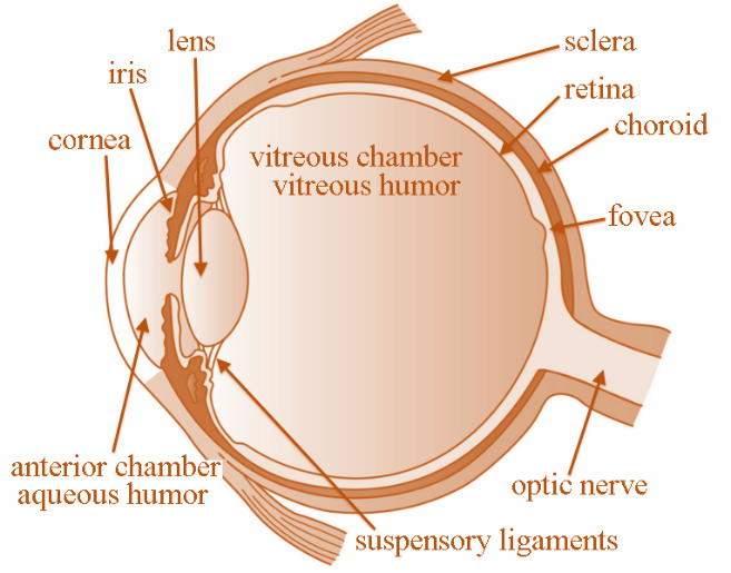
Humans have a pair of eyes located in cavities of the skull called orbits. The part of the eye that is visible to us comprises the cornea, sclera, iris, and pupil. The sclera is covered by a very thin layer called conjunctiva. The white visible part of the eye is called cornea – it is transparent and curved. The pupil is an aperture like structure located at the center of the iris.
The posterior part of the eye comprises the lens, retina, aqueous and vitreous humour and the optical nerve.

Fig: Structure of the Human Eye.
Eye contains the photoreceptor cells called rod cells and cone cells. These cells are present in the retina. Light falling on the eye is channeled to these photoreceptors by the pupil. The light energy is then converted to electrical signals and transmitted to the brain. The electric signals are interpreted as sight or vision.
The rod cells are responsible for night vision, while the cone cells for day vision or bright light vision. These cells contain a photopigment called Rhodopsin. Rhodopsin in turns comprises a protein called opsin and a light absorbing molecule called retinal.
The mechanism of vision can be explained in the following steps:
Light rays in the visible spectrum are focused on the retina with the help of cornea and lens.
This generates an impulse that causes dissociation of rhodopsin into opsin and retinal.
This dissociation in response to light, causes alteration in the structure of opsin.
The change in opsin structure leads to changes in the permeability of membranes of the eye which lead to potential differences in the photoreceptor cells.
The photoreceptors are connected to the bipolar cells through synapses. The bipolar cells in turn form synapse with the ganglion cells. Therefore, potential difference generated in the photoreceptor causes the ganglion cells to generate impulses.
The impulses are then transmitted to the visual cortex of the brain through the optical nerve.
The visual cortex analyzes the impulse which enables us to recognize the image formed on the retina and results in vision.
Note:- Human eye is the only photoreceptor sense organ in the body. It is composed of photoreceptor cells – the rods and cones. These cells can transduce light of the visible spectrum and send impulses to the brain, which results in vision. The light rays are focussed on the retina of the eye, that lead to formation of impulses in the rod cells and cone cells which, when transmitted to the brain, leads to image formation.
Complete step-by-step solution:-
Eye is the sense organ that is responsible for vision in animals. All the physiological components of the body responsible for vision are known as the visual system. This system is responsible for colour vision, motion perception, pattern recognition, sense of light.
In this section we will discuss the mechanism of vision in humans. To understand the mechanism of vision, first we have to understand the basic structure of the human eye.
Humans have a pair of eyes located in cavities of the skull called orbits. The part of the eye that is visible to us comprises the cornea, sclera, iris, and pupil. The sclera is covered by a very thin layer called conjunctiva. The white visible part of the eye is called cornea – it is transparent and curved. The pupil is an aperture like structure located at the center of the iris.
The posterior part of the eye comprises the lens, retina, aqueous and vitreous humour and the optical nerve.

Fig: Structure of the Human Eye.
Eye contains the photoreceptor cells called rod cells and cone cells. These cells are present in the retina. Light falling on the eye is channeled to these photoreceptors by the pupil. The light energy is then converted to electrical signals and transmitted to the brain. The electric signals are interpreted as sight or vision.
The rod cells are responsible for night vision, while the cone cells for day vision or bright light vision. These cells contain a photopigment called Rhodopsin. Rhodopsin in turns comprises a protein called opsin and a light absorbing molecule called retinal.
The mechanism of vision can be explained in the following steps:
Light rays in the visible spectrum are focused on the retina with the help of cornea and lens.
This generates an impulse that causes dissociation of rhodopsin into opsin and retinal.
This dissociation in response to light, causes alteration in the structure of opsin.
The change in opsin structure leads to changes in the permeability of membranes of the eye which lead to potential differences in the photoreceptor cells.
The photoreceptors are connected to the bipolar cells through synapses. The bipolar cells in turn form synapse with the ganglion cells. Therefore, potential difference generated in the photoreceptor causes the ganglion cells to generate impulses.
The impulses are then transmitted to the visual cortex of the brain through the optical nerve.
The visual cortex analyzes the impulse which enables us to recognize the image formed on the retina and results in vision.
Note:- Human eye is the only photoreceptor sense organ in the body. It is composed of photoreceptor cells – the rods and cones. These cells can transduce light of the visible spectrum and send impulses to the brain, which results in vision. The light rays are focussed on the retina of the eye, that lead to formation of impulses in the rod cells and cone cells which, when transmitted to the brain, leads to image formation.
Recently Updated Pages
Master Class 12 Economics: Engaging Questions & Answers for Success

Master Class 12 Physics: Engaging Questions & Answers for Success

Master Class 12 English: Engaging Questions & Answers for Success

Master Class 12 Social Science: Engaging Questions & Answers for Success

Master Class 12 Maths: Engaging Questions & Answers for Success

Master Class 12 Business Studies: Engaging Questions & Answers for Success

Trending doubts
Which are the Top 10 Largest Countries of the World?

What are the major means of transport Explain each class 12 social science CBSE

Draw a labelled sketch of the human eye class 12 physics CBSE

Why cannot DNA pass through cell membranes class 12 biology CBSE

Differentiate between insitu conservation and exsitu class 12 biology CBSE

Draw a neat and well labeled diagram of TS of ovary class 12 biology CBSE


