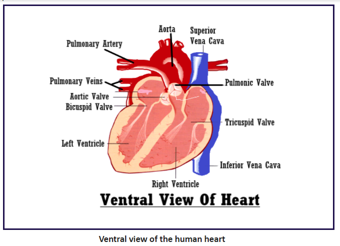
Sketch and label the ventral view of the human heart.
Answer
588.9k+ views
Hint: Human heart is divided into four chambers-two auricles and two ventricles. The wall of the ventricle is thicker than the wall of the auricle as it has to pump blood out of the heart with high pressure.
Complete answer:
The human heart is a highly muscular structure made of the cardiac muscle that pumps blood through the network of arteries and veins called the cardiovascular system. It is located just behind and slightly left of the breastbone. The ventral view or ventral side of the heart includes the superior vena cava, inferior vena cava, aorta, right ventricle, left ventricle, pulmonary artery, and the coronary artery. When the right auricle relaxes, deoxygenated blood from the superior and inferior vena cava pour into the right ventricle. When the auricles contract the deoxygenated blood moves through the atrioventricular valve (tricuspid) into the right ventricle while the oxygenated blood moves through the atrioventricular valve (bicuspid) into the left ventricle. The right ventricle after receiving deoxygenated blood transfers it into the pulmonary artery which transports it to the lungs for purification. Through the aorta, the left ventricle pumps blood to all parts of the body.
The sinoatrial node (SA node) is a myocardial structure where the electrical impulses stimulate contraction. The atrioventricular node (AV node) is a small mass of muscular fibers at the base of the wall between the atria.

Note: -Tricuspid and bicuspid valves are present between the right auricle and the right ventricle and also between the left auricle and left ventricle, to prevent the backflow of blood.
-The average human heart is the size of a fist in an adult and it weighs about 200 to 425 gms. It will beat about 115,000 times each day and pumps about 2,000 gallons of blood every day.
-An electrical system controls the rhythm of your heart. It’s called the cardiac conduction system. When the heart is disconnected from the body it can continue beating.
-The SA (sinoatrial) node generates an electrical signal that causes the contraction of chambers of the upper heart (atria). The signal then passes through the AV (atrioventricular) node to the chambers of the lower heart (ventricles), causing contraction there, for pumping.
Complete answer:
The human heart is a highly muscular structure made of the cardiac muscle that pumps blood through the network of arteries and veins called the cardiovascular system. It is located just behind and slightly left of the breastbone. The ventral view or ventral side of the heart includes the superior vena cava, inferior vena cava, aorta, right ventricle, left ventricle, pulmonary artery, and the coronary artery. When the right auricle relaxes, deoxygenated blood from the superior and inferior vena cava pour into the right ventricle. When the auricles contract the deoxygenated blood moves through the atrioventricular valve (tricuspid) into the right ventricle while the oxygenated blood moves through the atrioventricular valve (bicuspid) into the left ventricle. The right ventricle after receiving deoxygenated blood transfers it into the pulmonary artery which transports it to the lungs for purification. Through the aorta, the left ventricle pumps blood to all parts of the body.
The sinoatrial node (SA node) is a myocardial structure where the electrical impulses stimulate contraction. The atrioventricular node (AV node) is a small mass of muscular fibers at the base of the wall between the atria.

Note: -Tricuspid and bicuspid valves are present between the right auricle and the right ventricle and also between the left auricle and left ventricle, to prevent the backflow of blood.
-The average human heart is the size of a fist in an adult and it weighs about 200 to 425 gms. It will beat about 115,000 times each day and pumps about 2,000 gallons of blood every day.
-An electrical system controls the rhythm of your heart. It’s called the cardiac conduction system. When the heart is disconnected from the body it can continue beating.
-The SA (sinoatrial) node generates an electrical signal that causes the contraction of chambers of the upper heart (atria). The signal then passes through the AV (atrioventricular) node to the chambers of the lower heart (ventricles), causing contraction there, for pumping.
Recently Updated Pages
Master Class 12 Economics: Engaging Questions & Answers for Success

Master Class 12 Physics: Engaging Questions & Answers for Success

Master Class 12 English: Engaging Questions & Answers for Success

Master Class 12 Social Science: Engaging Questions & Answers for Success

Master Class 12 Maths: Engaging Questions & Answers for Success

Master Class 12 Business Studies: Engaging Questions & Answers for Success

Trending doubts
Which are the Top 10 Largest Countries of the World?

What are the major means of transport Explain each class 12 social science CBSE

Draw a labelled sketch of the human eye class 12 physics CBSE

What is a transformer Explain the principle construction class 12 physics CBSE

Why cannot DNA pass through cell membranes class 12 biology CBSE

Differentiate between insitu conservation and exsitu class 12 biology CBSE


