Courses
Courses for Kids
Free study material
Offline Centres
More
Store Icon
Store

Neural Control and Coordination Class 11 Notes CBSE Biology Chapter 18 [Free PDF Download]

ffImage
widget title icon
Latest Updates

widget icon
Enrol Now :
NEET Test Series
widget icon
Grab Your Seat :
NEET Pro Course
widget icon
Register Today :
NEET One to One Coaching

Revision Notes for CBSE Class 11 Biology Chapter 1 (Neural Control and Coordination) - Free PDF Download

Vedantu has created notes and short key-notes for the chapter, Neural Control and Coordination for Class 11, with the help of subject matter experts. This chapter gives insights about the entire neural system of all animals and their coordination. This is a very important chapter in Biology and will help you with higher studies. Vedantu has very experienced teachers to give you a better understanding of the subject. You can download the pdf of the notes from Vedantu’s website (Vedantu.com). The reference notes will definitely help you prepare for your exams.


  • The Nervous system coordinates communication within the body.

  • When two or more organs interact and complement the functions of one other by a process is known as coordination. For quick coordination, our neural system is organized into point-to-point connections.

  • The basic unit of neural organization is the neuron that detects, receives and transmits different stimuli.


Download CBSE Class 11 Biology Revision Notes 2024-25 PDF

Also, check CBSE Class 11 Biology revision notes for other chapters:


Important Chapter Wise Related Links

Competitive Exams after 12th Science
tp-imag
bottom-arrow
tp-imag
bottom-arrow
tp-imag
bottom-arrow
tp-imag
bottom-arrow
tp-imag
bottom-arrow
tp-imag
bottom-arrow

Neural Control and Coordination Class 11 Notes Biology - Basic Subjective Questions


Section–A (1 Mark Questions)

1. How many pairs of cranial nerves are present in man?

Ans. 12 pairs of cranial nerves are present in man.


2. Name the band of nerve fibres that joins the two cerebral hemispheres in mammals.

Ans. Corpus callosum is the band of nerve fibres that join the two cerebral hemispheres in mammals.


3. Rearrange the following in the correct order of involvement in electrical impulse movement-   Synaptic knob, dendrites, cell body, Axon terminal, Axon.

Ans. Synaptic knob → Dendrites → Cell body → Axon → Axon terminal. 


4. What is the function ascribed to Eustachian tube?

Ans. Eustachian tube carries extra sound in tympanic cavity to the pharynx. It is done to maintain optimum pressure around ear drum.


5. Name the fluid that surrounds and protects the brain. 

Ans. Cerebrospinal fluid surrounds and protects the brain.


Section–B (2 Mark Questions)

6. Define neurotransmitter.

Ans. Neurotransmitter is a chemical substance that acts as a signalling molecule which is released at the end of nerve fibre. It diffuses across the synapse and transmits the impulse to the next neuron, muscle or other structure.


7. The flowchart given here shows functional organization of the human neural system.

Identify A to E.
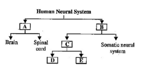

seo images


Ans.  


Nervous System


8. Complete the following paragraph by selecting the option that gives the correct sequence of words.

When a stimulus is applied at a site on the polarised membrane, the membrane at that site becomes freely permeable to __(i)__ ions. It causes rapid influx of __(ii)__ ions leading __(iii)__ of the membrane.

Ans. (i) Sodium ions.

(ii) Sodium ions

(iii) Depolarization 


9. Answer the following:

(a) Which part of the human brain is the most developed?

(b) Which part of our central neural system acts as a master clock?

Ans. (a) Forebrain is the largest and the most developed part of the human brain.

(b) Hypothalamus acts as a master clock in the human body.


10. What happens when the membrane of a nerve cell carries out a sodium pump?

Ans. When the membrane of a nerve cell carries out a sodium pump, it transports 3 Na+ ions outwards and 2 K+ ions into the cell.  As a result, the outer surface of the axonal membrane becomes a positively charge while its inner surface is negatively charged. It restores its resting membrane potential.


11. Name the meninges that surround the brain from outside to inside.

Ans. Dura mater

Arachnoid Layer

Pia mater


PDF Summary - Class 11 Biology Neural Control and Coordination Notes (Chapter 18)


Neural Control and Coordination

The interaction of two or more organs among each other and to complement each other’s function is known as coordination.

Neural System

The structural and functional units of the neural system are known as neurons. They can detect, receive, and transmit stimuli.

Human Nervous System

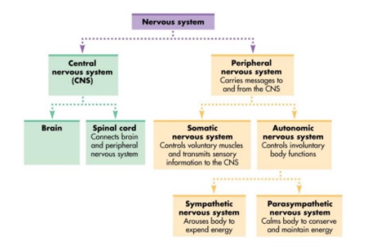
The classification of the nervous system is done into two types- 

(i) central nervous system (CNS) 

(ii) peripheral nervous system (PNS). 

The brain and Spinal cord are the 2 parts of the Central nervous system whereas the peripheral nervous system is composed of all the nerves present in the brain and the spinal cord. The two types of nerve fibers present in PNS are- 

(a) afferent nerve fibers or sensory nerve fiber

(b) efferent nerve fibers or motor nerve fiber

Impulses are carried from organs/tissues to the CNS in Afferent nerve fibers whereas, in efferent nerve fibers, impulses are transmitted from CNS to the target tissue/organ. 

PNS Is Classified Into Two Types-

(i) somatic nervous system 

(ii) autonomic nervous system. 

Impulses are transmitted from the CNS to skeletal muscles in the somatic nervous system while the autonomic nervous system innervates cardiac and smooth muscles of the glands which transmit the impulses from the CNS to the involuntary part of the body. The classification of the autonomic nervous system is done into two types-

(i) sympathetic nervous system 

(ii) parasympathetic nervous system
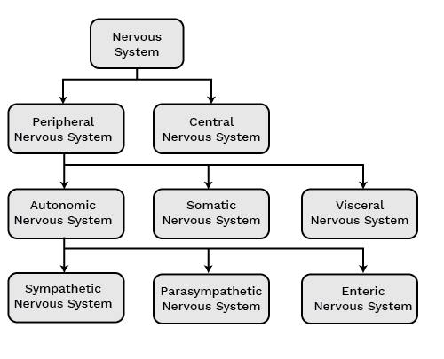

Types of Nervous Sysrem


Structure and the Function of the Neuron

A neuron is the structural and functional unit of the nervous system. It comprises 3 parts- 

(i) cell body 

(ii) dendrites  

(iii) axon. 

The cell body is made up of cytoplasm that contains granular bodies called Nissl’s granules. Short, several, finger-like projections that arise from the cell body are called dendrites. Dendrites carry nerve impulses towards or into the cell body. Axon is a long, single structure whose distal end is branched. Each of the axons ends into a bulb-like structure known as the synaptic knob, which contains neurotransmitters. The cells of the axon which produce and cover the myelin sheath are known as Schwann cells. The interruption between the two-adjacent myelin sheath is known as Nodes of Ranvier.
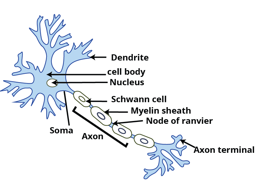

Structure of of the Neuron


Types of Neurons

Multipolar Neurons- Several processes arise in them, having one axon and many dendrites. For example, in the cerebral cortex.

Bipolar Neurons- They are the ones with one axon and one dendrite. For example, retina.

Unipolar Neurons- Unipolar neurons have only two fibers arising close together, i.e., one axon, no dendrite. For example, embryonic stage. 
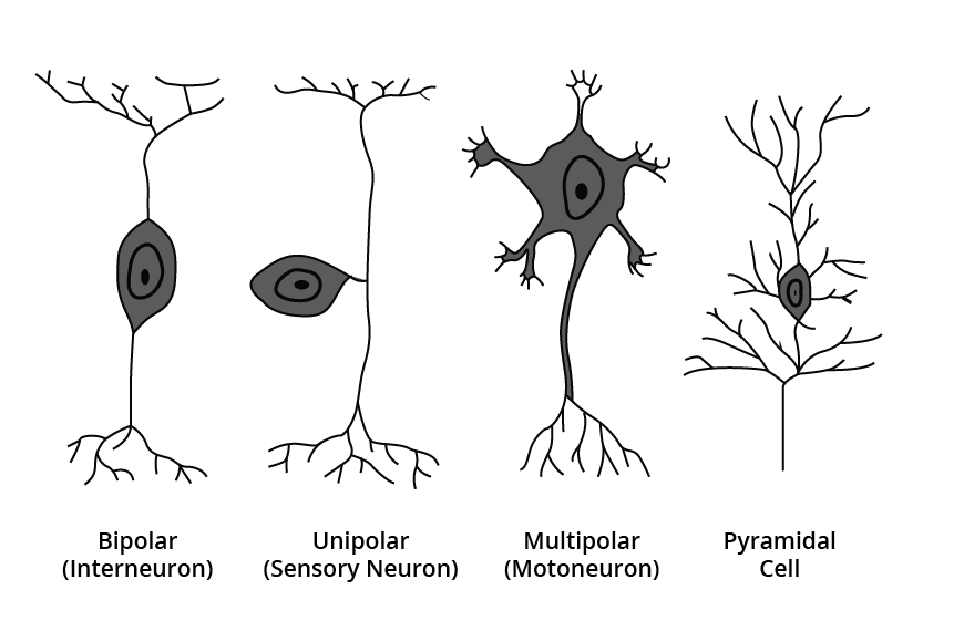

Types of Neuron


Generation and Conduction of Nerve Impulses

The cells which can be stimulated and electrically excited are called neurons. The membrane of the neurons is in a polarized state. The membrane contains selectively permeable ion channels with varying permeability to different ions. The membrane becomes more permeable to potassium ions and impermeable to sodium ions when no nerve impulse is being transmitted. This is the resting membrane potential. Therefore, there is a high concentration of potassium ions present in the membrane as compared to sodium ions. But, the case becomes opposite outside, high concentration of potassium in comparison to sodium. A concentration gradient gets developed due to the difference in the concentration of these ions. Sodium potassium pump helps in maintaining this gradient by actively transporting 3 sodium ions outside and 2 potassium ions inside the neuronal membrane. Thus, the outer surface of the axonal membrane possesses a positive charge and its inner surface becomes negatively charged. This is the reason behind the depolarization of the membrane. The membrane becomes freely permeable to sodium ions when a stimulus reaches a neuronal membrane. Thus, it will cause an influx of sodium ions inside the neuronal membrane. The inner side of the membrane becomes positively charged whereas outside the membrane is negatively charged due to this polarity. An electrical potential difference gets created called action potential.
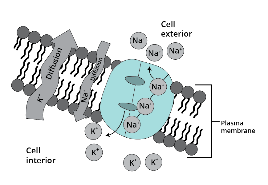

Action Potential


The permeability of sodium ions decreases, and the permeability of potassium ions increases with time. In the axonal membrane, there is an efflux of sodium ions polarization which occurs from inside to outside. Again, the polarity is reversed. This sequence of events is repeated along the complete axon which helps in the conduction of the nerve impulse.

Transmission of Impulses

The transmission of the nerve impulse from one neuron to another neuron via junctions called synapses. Synapse is a junction connecting the membrane of pre-synaptic neuron and postsynaptic neuron separated by a gap known as the synaptic cleft.

Two Types of Synapses are There- 

(i) Electrical synapse 

(ii) Chemical synapse. 

In an electrical synapse, there is proximity between pre-and post-synaptic neurons. There can be the direct flow of electric current from one neuron to another neuron. This mode of nerve impulse transmission is quicker than the chemical synapse.

In chemical synapses, pre-and post-synaptic neurons are separated by a fluid-filled space referred to as synaptic cleft. There are neurotransmitters involved in these synapses. There are axon terminals present which contain vesicles that are filled with neurotransmitters. By the time, the impulse reaches the axon terminal, synaptic vesicles start moving towards the membrane and fuse with it. This leads to the discharge of the neurotransmitters from the synaptic cleft. Now, the neurotransmitters bind with the receptors which are present on the post-synaptic neuron. Thus, ion channels get opened and it results in the transmission of fuse nerve impulses.
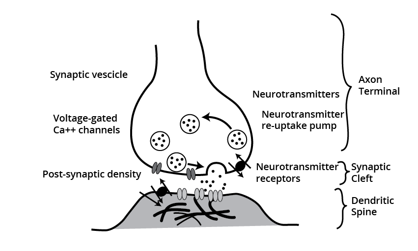

Transmission of Impulses


Central Nervous System

The brain controls and coordinates all the functions of the body thus, performing as a command and control system. The protection of the human brain is inside the skull. The covering of the brain is done in 3 layers known as meninges. They are-

(i) outermost dura mater, 

(ii) middle arachnoid, 

(iii) innermost pia mater. 

The division of the brain is into:

(a) forebrain, 

(b) midbrain, 

(c) hindbrain.

The forebrain consists of 3 parts- cerebrum, thalamus, and hypothalamus. The cerebrum is considered to be the largest part of the brain. The division of the cerebrum into two halves is known as the left and right cerebral hemispheres. These hemispheres are joined by a great white central canal white commissure called the corpus callosum. The covering of cerebral hemispheres by a layer of cells is known as the cerebral cortex. The thalamus is the area present at the center of the forebrain. It is involved in sensory and motor signaling. Hypothalamus lies at the base of the thalamus. The urge for eating, drink, and body temperature is all controlled and regulated by it.

The location of the midbrain is in between the forebrain and hindbrain. There are 4 lobes present known as Corpora quadrigemina. 

The parts of the hindbrain are- pons, cerebellum, and medulla oblongata. The cerebellum is considered to be the second-largest part of the brain. The medulla oblongata is placed between the spinal cord below and the pons above. The medulla controls gastric secretion, control respiration, and cardiovascular functions.
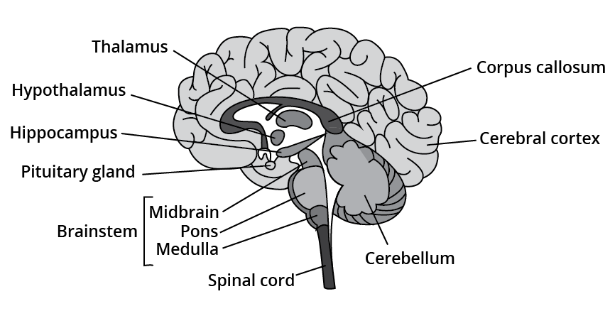

Central Nervous System


Reflex Action and Reflex Arc

Reflex action is an immediate involuntary stereotyped response. The neural elements involved in reflex action follow an arc called the reflex arc. Reflex arc or reflex pathway includes:

Receptor (sensory cell receiving stimulus)

   ↓

Afferent or sensory neuron

   ↓

The dorsal root of the spinal cord

    ↓

Connecting neuron in CNS

    ↓ 

Efferent or motor neuron

    ↓

The ventral root of the spinal cord

    ↓

Effector organ


Reflex Action and Reflex Arc


Sensory Perception and Processing

Sensory organs receive information from the environment in the form of mechanical, chemical, electrical, thermal and change it into nerve impulses which are transmitted to the central nervous system.

Human Eye

The human eye is spherical in structure, of which the anterior one-fifth is exposed while the four-fifth part remains concealed within the eye orbit. It is made up of 3 layers- 

 (i) the outermost sclera (dense connective tissue), 

 (ii) middle choroid (supplied with blood vessels),

 (iii) innermost retina.

Choroid lines the posterior region of the sclera. Anteriorly, the choroid enlarges to form the ciliary body. In front of the ciliary body, the choroid becomes widely separated from the cornea forming a visible colored portion of the eye known as the iris. A solid, transparent, crystalline biconvex lens is present which is just behind the pupil and is held by ligaments that are attached to the ciliary body.

There are 3 layers of neuronal cells in the Retina- 

(i) Ganglion cells,

(ii) Bipolar cells, 

(iii) Photoreceptor cells. 

There are two types of photoreceptor cells, Rods, and Cones. They are light-sensitive proteins. Cones are sensitive to the bright light of high intensity and color vision whereas rods are more sensitive to dim light. The protein present in Rods is known as rhodopsin. Cones particularly respond to 3 different colors- red, green, and blue.


Layers of neuronal cells in the Retina


The nerve connecting the eye to the brain is the optic. There is an absence of photoreceptors in this region, hence it is called a blind spot. Macula lutea is present which is lateral to the blind spot which is densely packed with only cones. The aqueous chamber is a fluid-filled space present in the cornea between the lens and fluid known as an aqueous chamber and fluid is called aqueous humor. Consequently, the space between the lens and the retina is known as the vitreous chamber and the fluid in it is known as vitreous humor.

Mechanism of Vision

An action potential gets generated in rods and cones when light rays are focused on the retina. Because of this, a photosensitive pigment gets separated into opsin (protein component) and retinal (aldehyde of vitamin A). This helps in generating an action potential that travels from the optic nerve to the visual cortex of the brain. This results in the formation of images on the retina.

The Ear

There are two main functions of the ears, hearing and balancing. There are 3 parts of the ear: 

(i) outer ear, 

(ii) middle ear, 

(iii) inner ear. 

Pinna and external auditory meatus are the two parts of the Outer ear. Pinna helps in the production of sound by collecting the vibration from the ear. Wax secreting glands are present in pinna and meatus.

The 3 ossicles of middle ear are: malleus, incus and stapes. The middle ear is connected to the pharynx by a eustachian tube. The inner ear comprises the bony labyrinth that contains 3 semicircular canals. The coiled part of the labyrinth is known as the cochlea. T Endolymph is the space filled within the cochlea. Structure located on the basilar membrane is known as the organ of Corti. They contain hair cells that act as auditory receptors. 


The Ear


Mechanism of Hearing 

The external ear receives sound waves and transfers them to the eardrum. This, in turn, vibrates the eardrum which transfers the vibrations to the ear ossicles. The vibrations are further transferred to the cochlea. This generates the nerve impulse which is transmitted via the auditory nerve to the auditory cortex of the brain. 

Human Neural System

The human neural system is composed of -

(i). Central Neural System (CNS)

(ii). Peripheral Neural System (PNS)

The CNS comprises the brain and the spinal cord and is the place of information processing and control. The PNS includes all the nerves of the body linked with the CNS. 

The nerve fibres of Peripheral Neural System (PNS) are of two types:

(a). Afferent Fibers: They carry impulse from tissue organs to the CNS.

(b). Efferent Fibers: They transmit impulse from the Central Neural System to the peripheral organ’s tissue. 

There are two divisions in the Peripheral nervous system. 

                                      Peripheral Nervous System


          ↓

Somatic Nervous System         Autonomic Nervous System

( it relays impulse from         (it transmit impulse from CNS 

CNS to skeletal muscle)           to the muscles of the body)

The Autonomic Nervous system is further divided.

                                      Autonomic Nervous System

_______________________________________________________________

                           ↓             ↓

Sympathetic neural system parasympathetic neural system

Note: The Autonomic Nervous System is also called visceral nervous system, which is also a part of PNC. The visceral nervous system includes nerves, fibres, ganglia and plexuses through which impulses travel from the CNS to the viscera and from the viscera to the CNS.

A neuron is the basic structural and functional unit of the nervous system.

It comprises mainly three major parts namely cells body, dendrites axon. 

(a). Cell Body: Cell body has cytoplasm with cell organelles and granular bodies that are also called Nissl’s granules.

(b). Dendrites: They are short fibres, which branch repeatedly, and projects out of the cell body. They too contain Nissl’s granules. These fibres transmit impulses towards the cell body. 

(c). Axon: It is a long fibre whose distal end is branched. Each branch of the axon terminates as a bulb-like structure called synaptic knob. 

(d). The synaptic knob has synaptic vesicles that contain chemicals called neurotransmitters. 

The neurons are divided into three types on the basis of the number of axons and dendrites.

(a). Multipolar: This neuron has one axon and two or more dendrites. It is found in the cerebral cortex.

(b). Bipolar: This neuron has one axon and one dendrite. It is present in the retina of the eye.

(c). Unipolar: This neuron has a cell body with one axon only. It is found in the embryonic stage.

(Image Will Be Uploaded Soon)

(Image Will Be Uploaded Soon)

The two types of Axons are myelinated and nonmyelinated nerve fibres. 

  • The myelinated nerve fibres are sheathed with Schwann cells, which form a myelin sheath around the axon. 

  • There are gaps between the two adjacent myelin sheaths, which are called nodes of Ranvier. 

  • Schwann cells enclose the nonmyelinated nerve fibres that do not form a myelin sheath around the axon. They are present in spinal and cranial nerves.

Generation of Nerve Impulse

  • The neurons are excitable cells and get stimulated because their membranes are in a polarized state.

  • There are different types of ion channels present on the neural membrane. These ion channels are permeable to different ions selectively.

  • In the resting phase, the axonemal membrane is more permeable to K+ ions and less for Na+ ions.

  • K+ ions are more concentrated inside the axonemal membrane than outside, where Na+ ion concentration is more outside. The ionic gradient is maintained y Na+/ K+ pump. 

  • When the stimulus is given to the neurons become depolarized as Na+ ions move inside the membrane and polarity of the membrane is reversed.

  • The electrical potential difference across the plasma membrane is called action potential or a nerve impulse. This sequence is repeated along the length of the axon. 

  • The rise in the stimulus-induced permeability to sodium ions is extremely short-lived. It is quickly followed by the rise in permeability to potassium ion. The ions move outside the membrane and restore the resting potential of the membrane.

Transmission of Impulses

Synapse is a neuromuscular junction in which nerve impulse is transmitted from one neuron to another. It is formed by the transmission from one neuron to another. The membranes of a presynaptic neuron and postsynaptic neuron form it.

  1. The synaptic cleft is the gap between the pre-synaptic neuron and postsynaptic neuron. 

  2. Two types of Synapses are Chemical synapses and Electrical synapses.

  3. In electrical synapse pre and postsynaptic neurons are in close proximity. It flows directly from one neuron into another neuron. It is faster than the chemical synapse.

  4. Chemical synapse is reported by synaptic cleft.

  5. When an impulse or an action reaches the axon terminal, it stimulates the movement of the synaptic vesicle towards the membrane.

  6. These synaptic vesicles contain neurotransmitter synaptic vesicles. 

  7. Fuses with the plasma membrane and releases neurotransmitters in the synaptic cleft and the neurotransmitter that is released bind to the receptor present on postsynaptic membranes. This opens the ion channels and allows the entry of ions, which generates a new impulse in postsynaptic neurons. 

Central Nervous System

The central nervous system controls various voluntary and involuntary movements. 

  • Skull well protects the brain. Three membranes called cranial meninges cover the brain. These membranes are:

  1. Duramater ( it is the outer layer)

  2. Arachnoid (it is the middle layer)

  3. Piamater ( it is the inner layer)

  • The three major parts of the brain are:

  1. Forebrain

  2. Midbrain

  3. Hindbrain

Forebrain

The forebrain consists of Cerebrum, Thalamus and Hypothalamus

  • Cerebrum

  • Cerebrum is the largest part of the brain.

  • Cerebrum is divided longitudinally into two cerebral hemispheres, which are connected by the corpus callosum.

  • The cerebral hemisphere is covered by the cerebral cortex and it folds to form prominent folds.

  • Cerebral cortex is also known as grey matter and it contains the motor area and sensory area. 

  • The Association area performs complex functions like forming connections between sensory and motor areas. This is also known as white matter because they give an opaque white appearance to the layer.

  • Thalamus

  • The thalamus is wrapped up by the cerebrum. It regulates sensory and motor signalling. 

  • Hypothalamus 

  • It is present at the base of thalamus. It is the main centre that controls temperature and urges for eating and drinking. It also secretes hypothalamic hormones.

Midbrain

The midbrain is located between thalamus and hypothalamus.

  • Cerebral aqueduct passes through the midbrain.

  • The Dorsal portion consists of four lobes called corpora quadrigemina. Midbrain and hindbrain form the brainstem.

Hindbrain

Hindbrain comprises of 

  • Hindbrain consists of Pons that have fibre tracts.

  • Cerebellum has a convoluted surface to provide additional space for many neurons.

  • Medulla oblongata transmits signals to the spinal cord. It controls respiration, cardiovascular space reflexes and dastric decretions

(Image Will Be Uploaded Soon)

(Image Will Be Uploaded Soon)

Reflex action or Reflex Arc is the process of responding to the peripheral nervous system, which acts involuntarily.

  1. It comprises at least one afferent neuron or receptor and one efferent neuron.

  2. The reflex arc consists of afferent neurons that receive signals from sensory organs and transfer it to the CNS. Efferent neurons carry signals from CNS to the effectors.

(Image Will Be Uploaded Soon)

(Image Will Be Uploaded Soon)

Sensory Reception and Processing

  • The most valuable and sensitive sensory organs is the human eye, which enables us to see the wonderful world and colours around us. 

  • The eyes are known as visual receptors because they carry specialized receptor cells, which are sensitive to light.

  • The eyeball constitutes three specialized structures: sclera, choroid and retina.

  • The sclera of the eyeball is the white portion of the eye and maintains the shape of the eye.

  • The choroid layer is present behind the sclera and it absorbs scattered light and ensures a clear image on the retina. 

  • The choroid forms two structures: the iris and the ciliary body.

  • The dark muscular diaphragm is the iris, whose function is to control the size of the pupil. 

  • The function of the Pupil is to regulate the amount of light that reaches the retina.

  • The ciliary body is connected to the lens. It consists of smooth muscles and contraction of these muscles causes an alteration in the shape of the lens.

  • Lens is the transparent and flexible structure made up of protein. It creates an image on a light-sensitive screen called retina. A clear fluid that fills the lens with called aqueous humour. The back layer of the lens is filled with vitreous humour.

  • Light enters the eye through a transparent thin membrane called the cornea. 

  • Retina is the delicate third and innermost layer eye lens.  It receives light that the lens has focused and forms an inverted real image of the object. 

  • The retina has light-sensitive cells: cones and rods.

  • Cone cells contain a pigment called visual violet iodopsin that functions in the daylight and gives colour vision.

  • The rods cells function in dim light or night. 

  • When a person enters the darkroom from the lightroom, the person is not able to see properly for some time. It is because of the amount of light entering the eye and regulated by the iris. In light, the pupil contracts and permits less light to enter the eye. When a person enters the darkroom, the pupil takes some time to expand and allows more light to enter the eye. 

  • The minimum distance at which objects can be seen most clearly without strain also known as least distance of distinct vision is known as the Near point of an eye.

  • The point up to which the eye can see objects clearly is called the farthest point of the eye and it is infinity.

  • Cataract is the condition when the lens becomes milky and cloudy. It normally happens at an old age and it causes a partial or complete loss of vision.

Defect of Vision

The defects of vision are:

  1. Myopia: Myopia is caused due to excessive curvature of the eyeball. In Myopia, the image of the distant objects cannot be seen clearly. It is also called nearsightedness.

  2. Hypermetropia: In this condition, people can see faraway objects but cannot see nearby objects clearly. This is also called nearsightedness. Hypermetropia is caused due to the increase in the focal length and when the eyeball becomes small.

  3. Presbyopia: This defect is caused because of the gradual weakening of ciliary muscles and diminishing flexibility of the eye lens. 

(Image Will Be Uploaded Soon)

(Image Will Be Uploaded Soon)

  • Ear performs a very important function of hearing and maintenance of the body.

  • Ear is divided into three major sections: Outer ear, middle ear and inner ear.

  • Outer Ear consists of:

  • Pinna

  • External Auditory Meatus called canola. These extend up further to the tympanic membrane or eardrum. 

  • Pinna collects the vibration in the air and collects sound. In pinna, wax screating sebaceous glands are present. 

The Middle Ear Consists of Three Ossicles.

  • Malleus is attached to the tympanic membrane.

  • Stapes is the third bone that is connected to the oval window of the cochlea.

  • Incus

  • The regulation of transmission of sound waves to the inner ear is increased by Ossicles.

  • Middle ear cavity is connected to the pharynx through the Eustachian tube. 

  • Labyrinth is the fluid-filled in the inner ear and it has two parts: the bony and membranous

  • Labyrinth is filled by a fluid called endolymph and the coiled portion of a labyrinth is the cochlea.

  • Bony labyrinth is divided into upper scala vestibuli and lower scala tympani. 

  • The basilar membrane organ of Corti is present, which possesses a hair cell, which acts as an auditory receptor. 

  • Stereocilia is a large number of processes that are projected from the apical part.

  • Inner Ear consists of a complex system called the vestibular apparatus. This apparatus is composed of three semicircular canals and the otolith organ. It consists of sacculi and utricle.

  • The base of the canals is swollen and is known as ampulla. It contains a projecting ridge called crista ampullaris. Macula is the projecting ridge between saccule and utricle. 

  • The external ear receives sound waves and directs them to the eardrum, which vibrates in response to the sound waves. These sound waves are transmitted through the ear ossicles. 

Sense Organs

The human body has five important senses: touch, vision, hearing, smell, and taste. These sensory receptors are present throughout the body; in the skin, mucous membranes, connective tissues and muscles. They provide various sensory signals like heat, cold, pain, etc. The sensory organs work together to accomplish a specific receptive process.

FAQs on Neural Control and Coordination Class 11 Notes CBSE Biology Chapter 18 [Free PDF Download]

1. What is the Function Ascribed to the Eustachian Tube?

A Eustachian tube connects the middle ear activity with the pharynx. The function of a Eustachian tube is to help in controlling the pressures on either side of the eardrum and equalizes the air pressure outside.

2. What is the Difference Between Afferent and Efferent Neurons?

The afferent nerve fibres transmit impulses from tissues or organs to the CNS whereas the efferent fibres transmit from the CNS to the concerned to the peripheral tissues or organs.

3. What is Aqueous Humour?

It is the space between the cornea and the lens and it is filled with a thin watery fluid. 

4. Name the Structure Involved in the Protection of the Brain.

The Skull well protects the brain. The cranial meninges cover the brain that consists of an outer layer called dura mater. Dura mater is a very thin middle layer called arachnoid and an inner layer called pia mater. 

5. What are the three main parts of the brain?

The brain is one of the most important and complex organs of our body. Our skull protects this important organ. Three main parts of the brain are: Cerebrum, Hypothalamus and Thalamus. Out of these three cerebrum is the most important part of the brain. Hypothalamus controls various functions of our body such as the release of hormones. Thalamus is the part of the brain that transmits information received by our senses to the cortex. 

6. What is the difference between the central neural system and the peripheral neural system?

The central neural system (CNS) comprises the brain and the spinal cord. On the other hand, the peripheral neural system (PNS) consists of the spinal and cranial nerves. There are no subdivisions in the CNS but the peripheral neural system is further subdivided into the autonomic nervous system and the somatic nervous system. PNS transmits impulses to the CNS which then processes the information and sends the responses to various parts of the body.

7. How to write good answers to score well in Chapter 18 of Class 11 Biology?

To score well in Chapter 18 of your Class 11 Biology exam, the most important thing is that the basics of this chapter must be strong and clear. Understand the pattern of the exam. Understand what the demand of the question is. Write your answer in simple language and use short sentences and appropriate terms. You should include appropriate diagrams, flow charts and tables in your answer. Make your answer look presentable and convey to the teacher that you know the concept.

8. How important is it to practice questions given in Class 11 Chapter 18?

Practising questions from Chapter 18 of Class 11 Biology provides you with an effective way to check your understanding. When you study a topic, you may forget it after some time, but if you practice the questions related to the topic, the information stays with you for a long time. Practising sufficient questions helps you find the right way to present your answers in the exam. It makes it easier for you to recall information in the exam and is an efficient way to revise what you have studied. To revise the chapter students can download the NCERT Notes for Class 11 Biology Chapter 18 free of cost from the vedantu website (vedantu.com).

9. What is given in Class 11 Biology Chapter 18?

The chapter explains to you the neural system of animals and their coordination. The nervous system enables communication within our mind and body. The neural system helps us detect and respond to various stimuli. The chapter differentiates between the Central Neural System and the Peripheral Neural System. It explains the basic structure of the nervous system and the functions of the organs involved. It also teaches you how your sensory organs function. To revise the chapter and know more about it students can download the vedantu app.