


Chemistry Notes for Chapter 10 Biomolecules Class 12 - FREE PDF Download
In Class 12 Chemistry, Biomolecules give insight into the essential organic compounds found in living organisms. This chapter covers the structure, functions, and properties of carbohydrates, proteins, lipids, and nucleic acids. Understanding these biomolecules is crucial for grasping biological processes and their significance in health and disease. This chapter provides insights into the molecular basis of life, laying the groundwork for more advanced studies in biochemistry and molecular biology.
 Table of Content
Class 12 Chapter 10 Biomolecules Notes lets you quickly access and review the chapter content. For a comprehensive study experience, check out the Class 12 Chemistry Revision Notes FREE PDF here and refer to the CBSE Class 12 Chemistry syllabus for detailed coverage. Vedantu's notes offer a focused, student-friendly approach, setting them apart from other resources and providing you with the best tools for success.
1. Introduction
Biomolecules are complex organic compounds that govern the common activities of living organisms. Carbohydrates, proteins, nucleic acids, lipids, and other complex biomolecules make up living systems. Furthermore, simple molecules such as vitamins and mineral salts play an important role in the functions of organisms.
2. Carbohydrates
Plants are the primary producers of carbohydrates, which comprise a large group of naturally occurring organic compounds. Cane sugar, glucose, starch, and other common examples The majority of them have the general formula \[{{\text{C}}_x}{{\text{H}}_{2y}}{{\text{O}}_y}\] and were thought to be carbon hydrates, hence the name carbohydrate. The molecular formula of glucose \[\left( {{{\text{C}}_{\text{6}}}{{\text{H}}_{{\text{12}}}}{{\text{O}}_{\text{6}}}} \right){\text{,}}\] for example, fits into this general formula, \[{{\text{C}}_{\text{6}}}\left( {{{\text{H}}_{\text{2}}}{\text{O}}} \right){\text{6}}{\text{.}}\] However, not all of the compounds that fit into this formula are carbohydrates. Rhamnose, \[{{\text{C}}_{\text{6}}}{{\text{H}}_{{\text{12}}}}{{\text{O}}_{\text{5}}}{\text{,}}\] is a carbohydrate, but it does not fall under this definition. Carbohydrates can be defined chemically as optically active polyhydroxy aldehydes or ketones, or compounds that produce such units upon hydrolysis. Some carbohydrates with a sweet taste are also known as sugars. Sucrose is the most common sugar found in our homes, whereas lactose is the sugar found in milk.
2.1 Classification of Carbohydrates
Carbohydrates are classified based on how they react to hydrolysis. They have been broadly classified into the three groups listed below:
2.1.1 Monosaccharides
A monosaccharide is a carbohydrate that cannot be hydrolyzed further to produce simpler units of polyhydroxy aldehyde or ketone. Examples include glucose, fructose, ribose, and others.
Monosaccharides are further classified based on the number of carbon atoms and the functional group they contain. When a monosaccharide contains an aldehyde group, it is referred to as an aldose, and when it contains a keto group, it is referred to as a ketose. As shown by the examples, the number of carbon atoms constituting the monosaccharide is also introduced in the name.
Different Types of Monosaccharides
2.1.2 Oligosaccharides
Oligosaccharides are carbohydrates that, when hydrolyzed, yield two to ten monosaccharide units. They are further classified as disaccharides, trisaccharides, tetrasaccharides, and so on, based on the number of monosaccharides produced during hydrolysis. Disaccharides are the most common of these. The two monosaccharide units formed by hydrolysis of a disaccharide may be identical or dissimilar. Sucrose, for example, yields one molecule of glucose and one molecule of fructose when hydrolyzed, whereas maltose yields only two molecules of glucose.
2.1.3 Polysaccharides
Polysaccharides are carbohydrates that when hydrolyzed yield a large number of monosaccharide units. Starch, cellulose, glycogen, gums, and other common examples of Polysaccharides do not have a sweet taste, so they are also known as non-sugars.
2.1.4 Reducing and Non-Reducing Sugars
Carbohydrates are also classified as reducing or non-reducing sugars. Reducing sugars are all carbohydrates that reduce Fehling's solution and Tollens' reagent. Sugars are reduced by all monosaccharides, whether aldose or ketose.
Nonreducing sugars, such as sucrose, are formed when the reducing groups of monosaccharides, such as aldehyde or ketonic groups, are bonded in disaccharides. Reducing sugars, on the other hand, are sugars that have these functional groups free, such as maltose and lactose.
3. Glucose (Aldohexose)
3.1 Preparation of Glucose
From Sucrose (Cane Sugar):
When sucrose is boiled in alcoholic solution with dilute \[{\text{HCl or }}{{\text{H}}_{\text{2}}}{\text{S}}{{\text{O}}_{\text{4}}}{\text{,}}\] glucose and fructose are obtained in equal amounts.
\[\mathop {{{\text{C}}_{{\text{12}}}}{{\text{H}}_{{\text{22}}}}{{\text{O}}_{{\text{11}}}}}\limits_{{\text{Sucrose}}} + {{\text{H}}_{\text{2}}}{\text{O}}\xrightarrow{{{{\text{H}}^ \oplus }}}\mathop {{{\text{C}}_{\text{6}}}{{\text{H}}_{{\text{12}}}}{{\text{O}}_{\text{6}}}}\limits_{{\text{Glucose}}} + \mathop {{{\text{C}}_{\text{6}}}{{\text{H}}_{{\text{12}}}}{{\text{O}}_{\text{6}}}}\limits_{{\text{Fructose}}} \]
From Starch
Glucose is commercially obtained by hydrolyzing starch with dilute \[{{\text{H}}_{\text{2}}}{\text{S}}{{\text{O}}_{\text{4}}}\] at 393 K under pressure.
\[\mathop {{{\text{C}}_{\text{6}}}{{\text{H}}_{10}}{{\text{O}}_{\text{5}}}}\limits_{{\text{Starch or cellulose}}} + n{{\text{H}}_{\text{2}}}{\text{O}}\xrightarrow[{393{\text{ K,2 - 3 atm}}}]{{{{\text{H}}^ \oplus }}}\mathop {{\text{n}}{{\text{C}}_{\text{6}}}{{\text{H}}_{{\text{12}}}}{{\text{O}}_{\text{6}}}}\limits_{{\text{Glucose}}} \]
3.2 Reactions
Oxidation
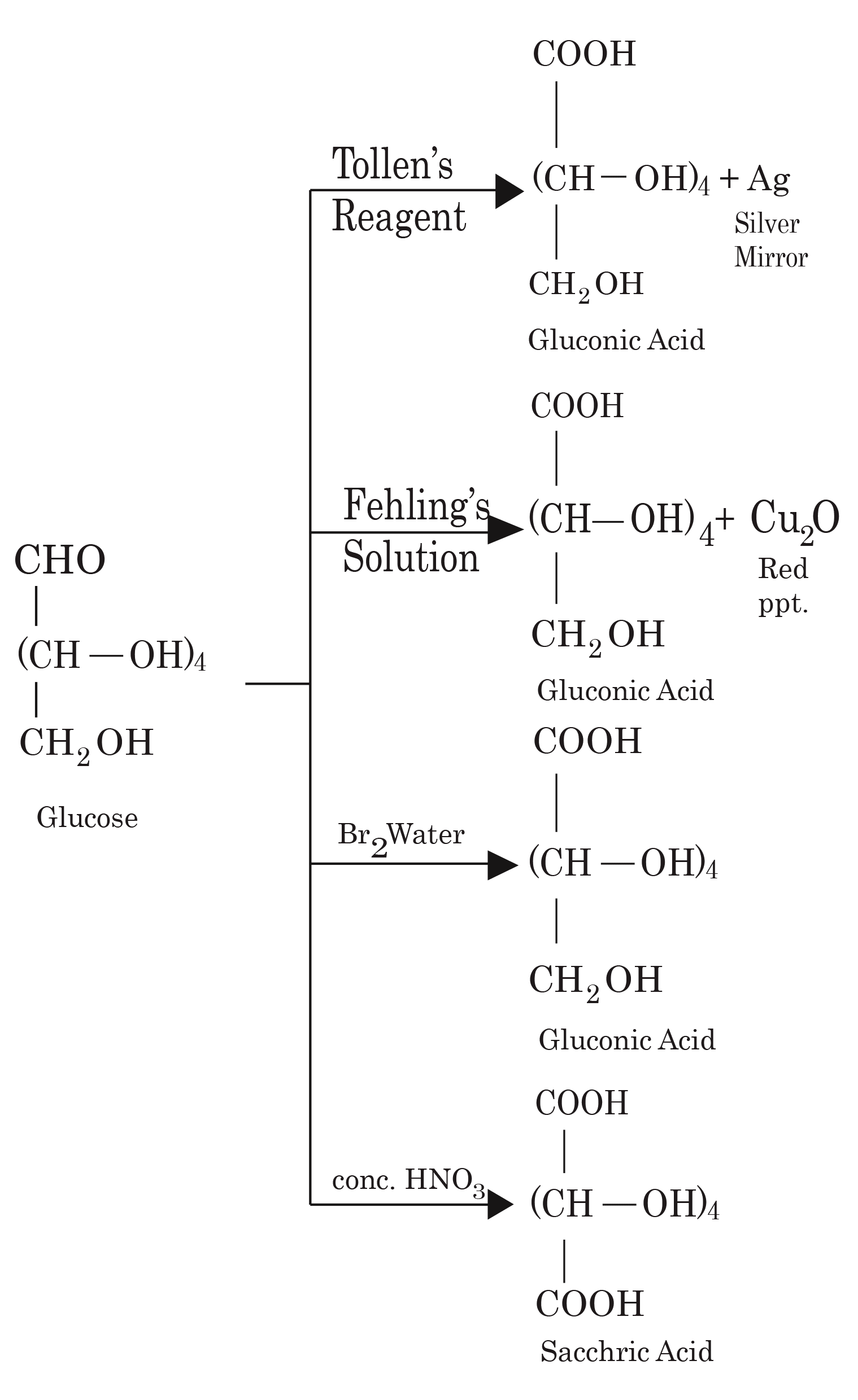
Oxidation Reactions of Glucose
Reduction
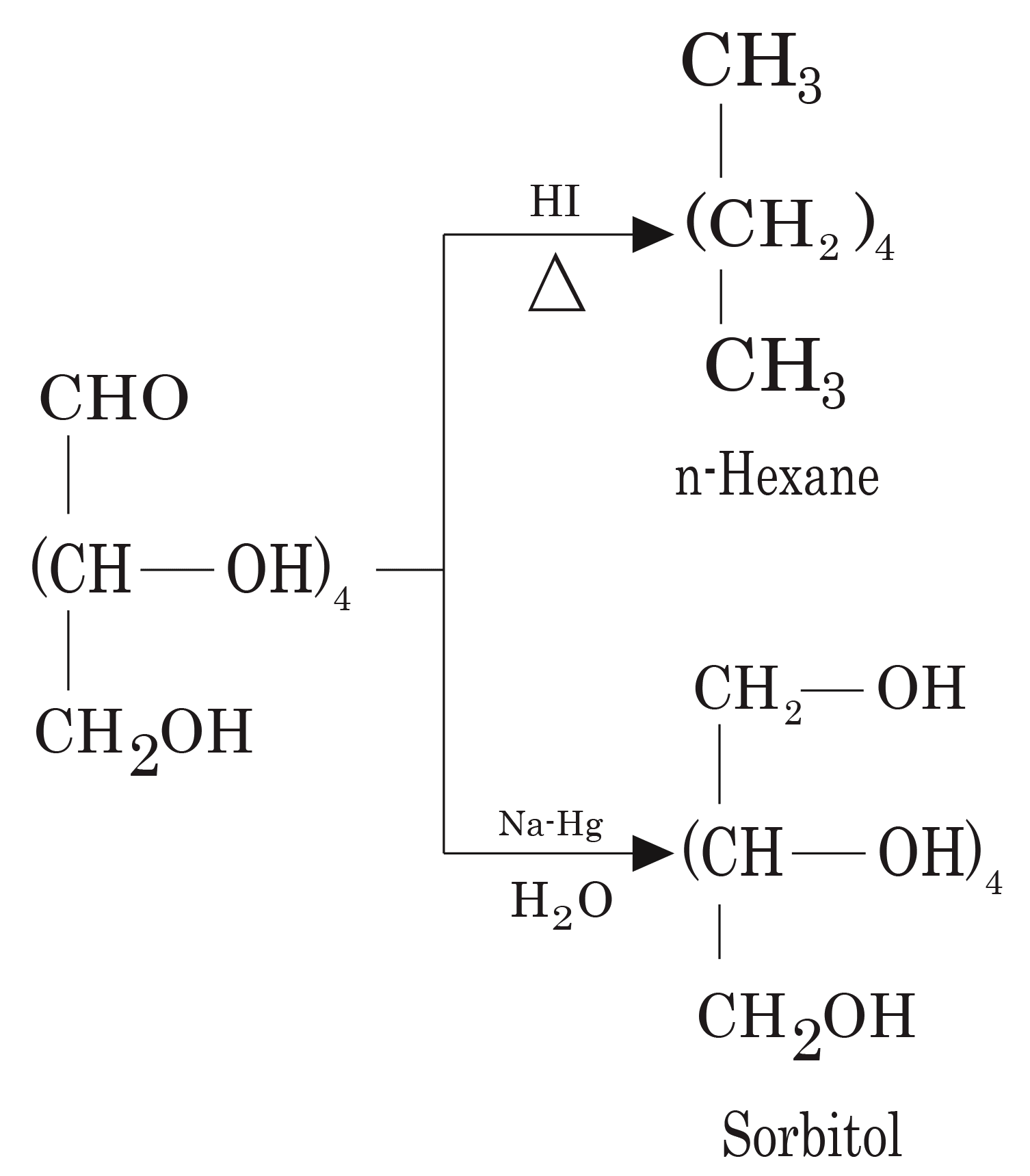
Reduction Reactions of Glucose
Note: Reduction with HI yields n-hexane, demonstrating that all six carbons of glucose are arranged in a straight chain.
Oxime Formation
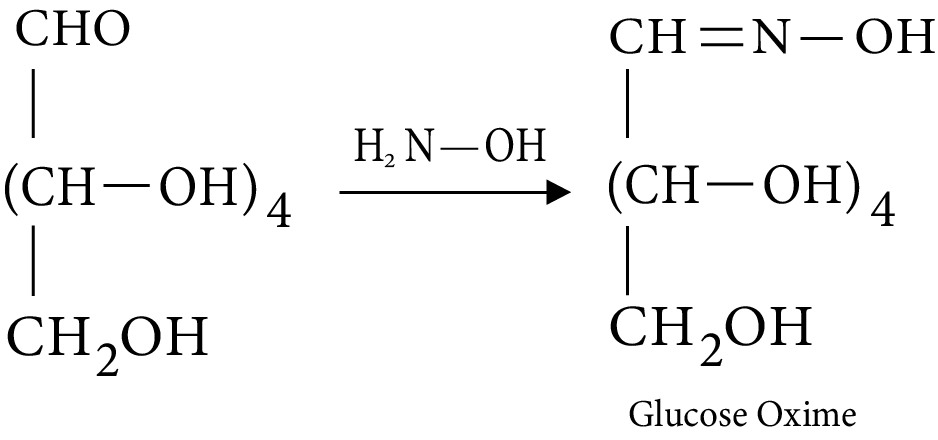
Oxime Formation by Glucose
Cyanohydrin Formation
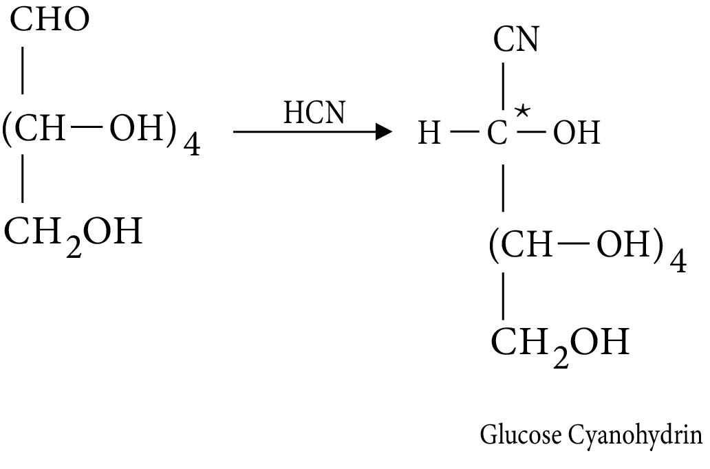
Formation by Glucose Cyanohydrin
Note: Since carbonyl C has become chiral, two diastereomers are formed.
Acetylation
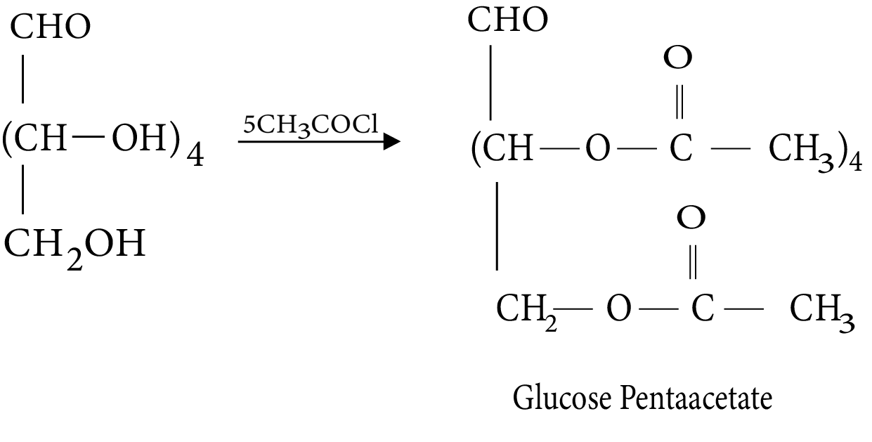
Acetylation of Glucose
Reaction with phenylhydrazine (formation of osazone)
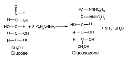
Reaction of Glucose with Phenylhydrazine
Note: To produce osazone, one mole consumes three moles of PhNHNH2. Two moles produce hydrazone, and one is used to convert a CHOH group to a carbonyl group.
3.3 Configuration in Monosaccharides
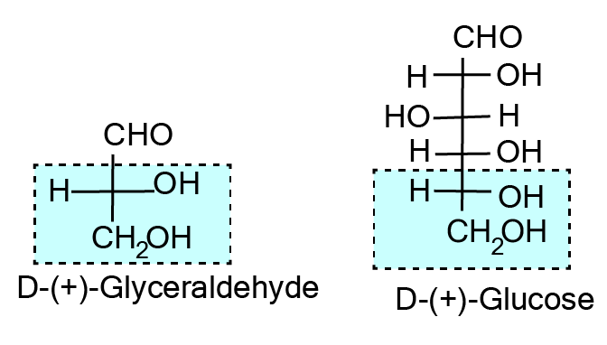
D(+)-glucose is the correct name for glucose. The letter ‘D' before the name of glucose represents the configuration, whereas the letter ‘(+)' represents the molecule's dextrorotatory nature. It should be noted that the letters 'D' and 'L' have no bearing on the compound's optical activity. The following is a definition of D– and L– notations. The letters 'D' or 'L' before the name of any compound indicate the stereoisomer's relative configuration. This refers to their affinity for a specific isomer of glyceraldehyde. Glyceraldehyde has one asymmetric carbon atom and comes in two enantiomeric forms, as shown.
Configuration in Glyceraldehyde
All compounds that can be chemically correlated to the (+) isomer of glyceraldehyde have D-configuration, whereas those that can be correlated to the (–) isomer of glyceraldehyde have L-configuration. The lowest asymmetric carbon atom (as shown below) is compared when assigning the configuration of monosaccharides. As with (+) glucose, –OH on the lowest asymmetric carbon is on the right side, similar to (+) glyceraldehyde, so it is assigned D-configuration. The structure is written in such a way that the most oxidised carbon is at the top for this comparison.
(+) Isomer of D-configuration in Glyceraldehyde and Glucose
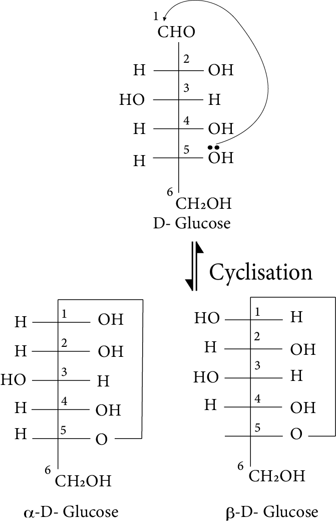
3.4 Cyclic Structure of Glucose
Glucose has been discovered to exist in two different crystalline forms, which are known as \[\alpha \]and \[\beta .\] The \[\alpha \]-form of glucose (m.p. 419 K) is obtained by crystallisation from a concentrated glucose solution at 303 K, whereas the \[\beta .\]-form (m.p. 423) is obtained by crystallisation from a hot and saturated aqueous solution at 371 K. In aqueous solution, both \[\alpha \]-D-glucose and \[\beta \]-D-glucose undergo mutarotation. Although the crystalline forms of \[\alpha \]- and \[\beta \]-D (+)-glucose are quite stable in aqueous solution, each form gradually transforms into an equilibrium mixture of the two.
This is demonstrated by the fact that the specific rotation of a freshly prepared aqueous solution of \[\alpha \]-D(+)-glucose decreases with time from \[{{ + 111^\circ to + 52}}{{.5^\circ ,}}\] whereas that of \[\beta \]-D(+)-glucose increases from \[{\text{ + 19}}{{.2^\circ to 52}}{{.5^\circ }}{\text{.}}\] Thus, mutarotation refers to the spontaneous change in specific rotation of an optically active compound with time to an equilibrium value. It was discovered that glucose forms a six-membered ring, with –OH at C-5 playing a role in ring formation. This explains the lack of a –CHO group as well as the presence of glucose in two forms, as shown below. These two cyclic forms coexist with an open chain structure.
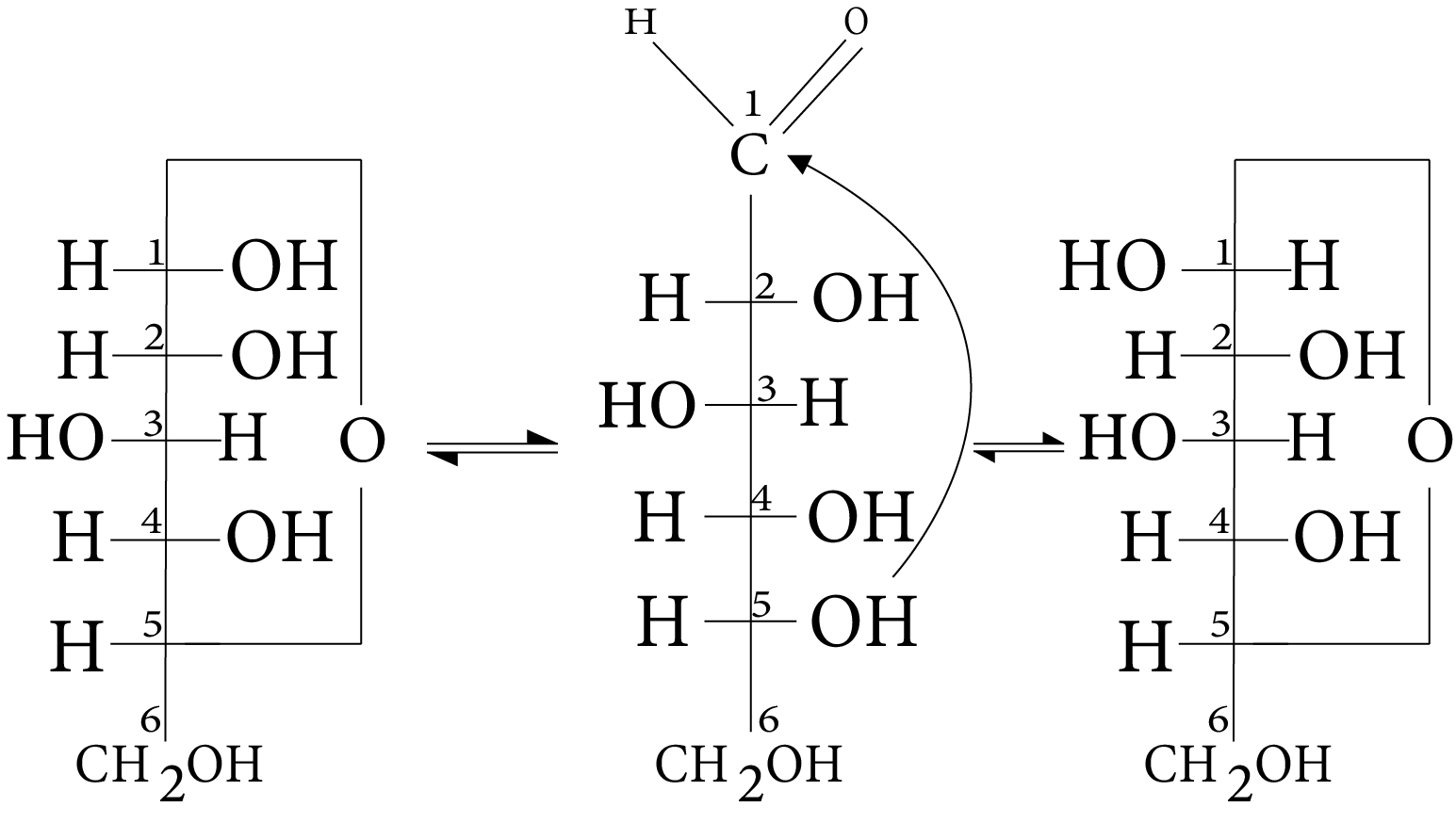
Cyclic Structure of Glucose
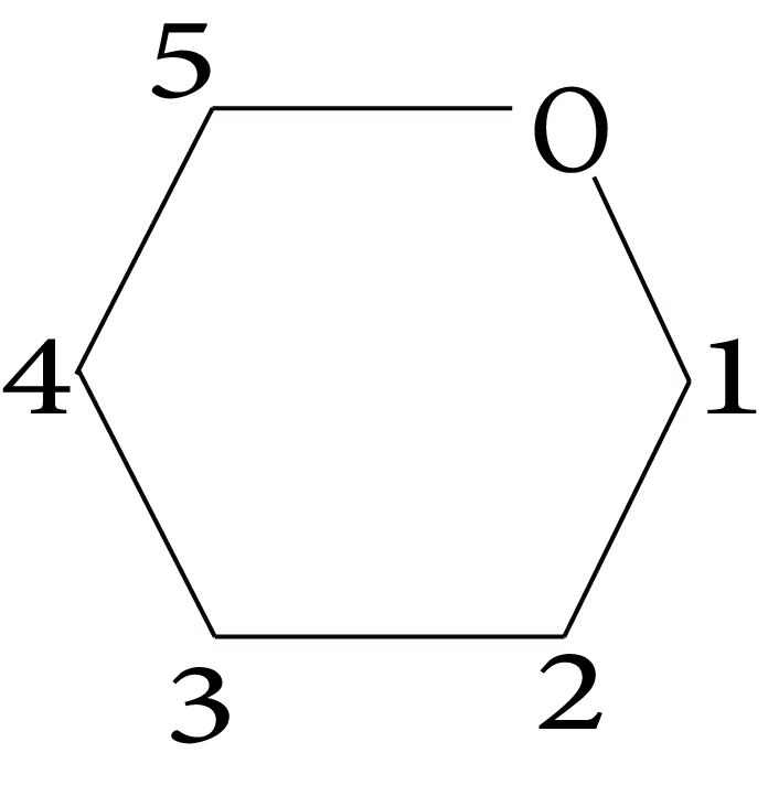
The only difference between the two cyclic hemiacetal forms of glucose is the configuration of the hydroxyl group at C1, which is referred to as anomeric carbon (the aldehyde carbon before cyclisation). Such isomers, namely -form and -form, are referred to as anomers. In analogy with pyran, the six-membered cyclic structure of glucose is known as the pyranose structure (– or –). Pyran is a cyclic organic compound with a ring containing one oxygen atom and five carbon atoms. The Haworth structure more accurately represents the cyclic structure of glucose.
Cyclic Structure of Glucose
3.4.1 How to Draw a Haworth Projection
A ring of 6 atoms (5 ‘C' and 1 ‘O') is drawn, with the ‘O' atom in the upper right hand corner, as shown below.
Numbering in the Haworth Projection
Number one is assigned to the carbon atom on the right side of oxygen. Then, in a clockwise fashion, other carbon atoms are assigned numbers 2, 3,........... . In Fischer projection, groups attached to a carbon on the right side are placed below the ring, while groups attached to the carbon on the left side are placed above the ring. However, by convention, the \[{\text{C}}{{\text{H}}_{\text{2}}}{\text{OH}}\] group of carbon 5 is placed above the plane of the ring.
(image will be uploaded soon)
4. Fructose (Ketohexose)
Fructose has the molecular formula \[{{\text{C}}_{\text{6}}}{{\text{H}}_{{\text{12}}}}{{\text{O}}_{\text{6}}}{\text{,}}\]and based on its reactions, it was discovered to have a ketonic functional group at carbon number 2 and six carbons in a straight chain, just like glucose. It is a levorotatory compound from the D-series. D-(–)-fructose is the correct spelling. Its open chain structure is depicted below.
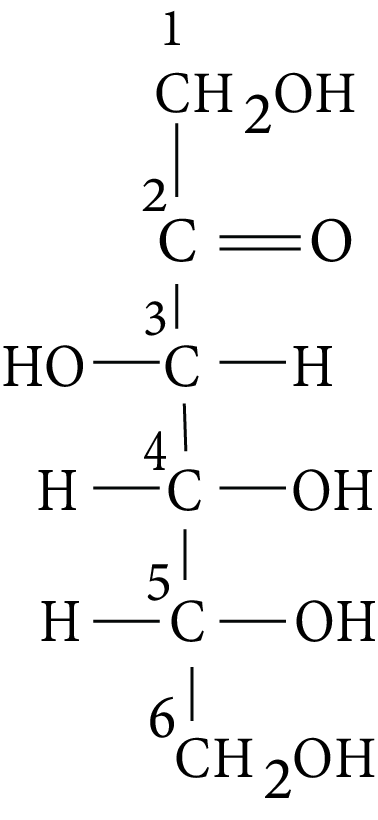
Fructose
It also exists in two cyclic forms, which are obtained by adding –OH to the carbonyl group at C5. The resulting ring is a five-membered ring known as furanose, after the compound furan. Furan is a five-membered cyclic compound that contains one oxygen atom and four carbon atoms.
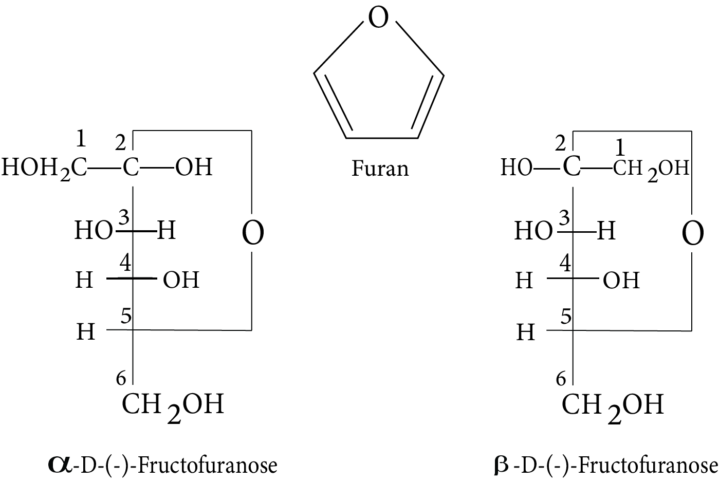
Anomers of Fructose
Haworth structures are used to represent the cyclic structures of two anomers of fructose.
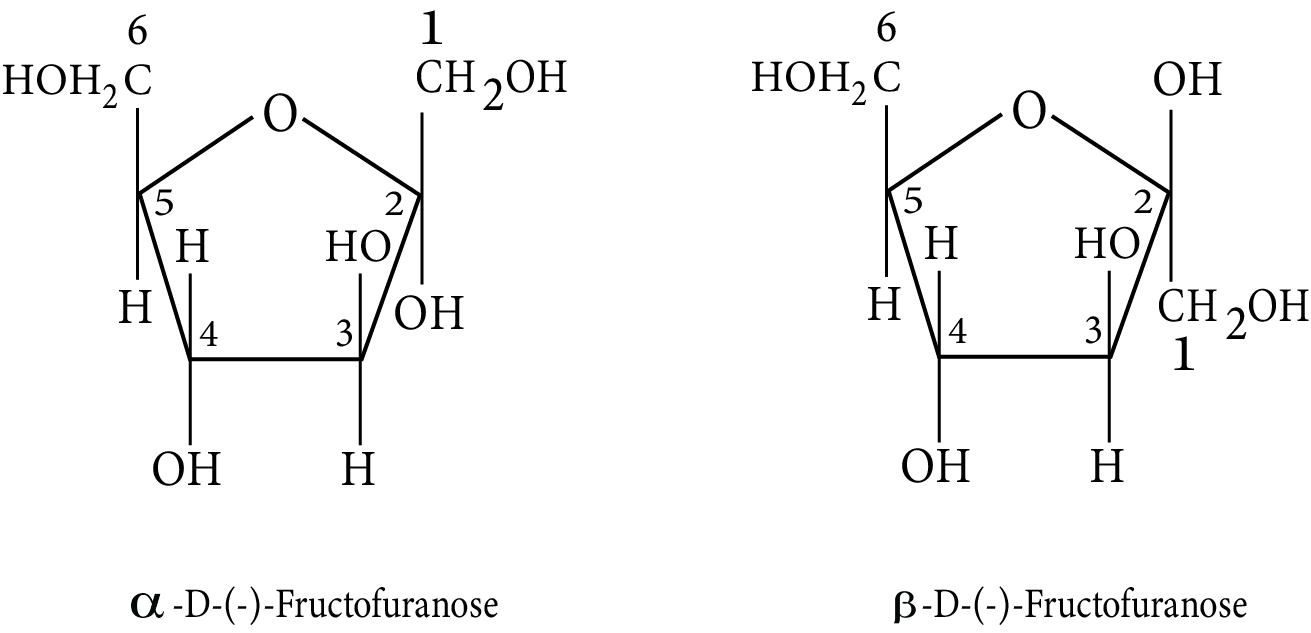
Haworth Structures of Anomers of Fructose
5. Comparison of Glucose And Fructose
6. Disaccharides
An oxide linkage formed by the loss of a water molecule connects the two monosaccharides. The linkage of two monosaccharide units via an oxygen atom is known as glycosidic linkage.
6.1 Sucrose
Sucrose is a common disaccharide that, when hydrolyzed, yields an equimolar mixture of D-(+)-glucose and D-(–)-fructose.
${\begin{matrix} C_{12}H_{22}O_{11} & + & H_{2}O \longrightarrow & C_{6}H_{12}O_{6} & + & C_{6}H_{12}O_{6} \\ Sucrose & & & D-(+)-Glucose & & D-(-)-Fructose \\ [\alpha]_D=+66.5^\circ & & & [\alpha]_D=+52.5^\circ & & [\alpha]_D=-92.4^\circ\end{matrix}} $
A glycosidic linkage between C1 of \[\alpha \]-glucose and C2 of \[\beta \]-fructose holds these two monosaccharides together. Sucrose is a non-reducing sugar because the reducing groups of glucose and fructose are involved in the formation of glycosidic bonds.
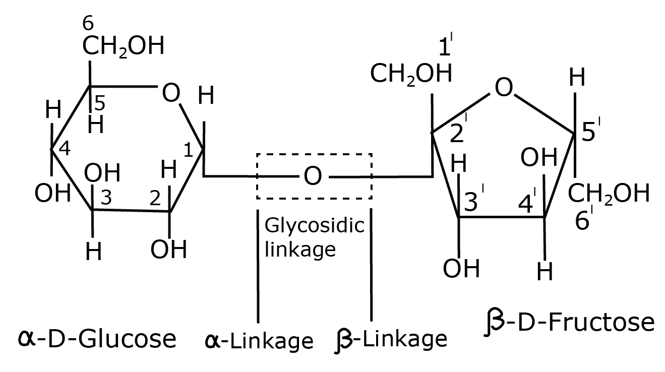
Sucrose
Sucrose is a dextrorotatory compound that when hydrolyzed yields an equimolar solution of glucose and fructose.
Because the laevo rotation of fructose is greater than the dextro rotation of glucose, this solution is laevorotatory.
Thus, hydrolysis of sucrose causes a change in the sign of rotation, from dextro (+) to laevo (–), and the product is known as invert sugar, and the phenomenon is known as sugar inversion.
6.2 Maltose
Maltose, another disaccharide, is made up of two -Dglucose units, with C1 of one glucose (I) linked to C4 of another glucose unit (II). Because the free aldehyde group can be produced at C1 of second glucose in solution and has reducing properties, it is classified as a reducing sugar.
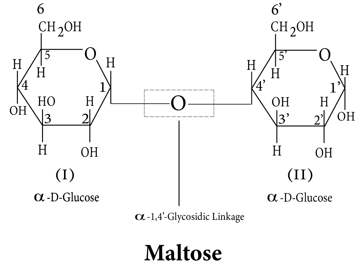
Maltose
6.3 Lactose
It is more commonly known as milk sugar since this disaccharide is found in milk. It is composed of \[\alpha \]-D-galactose and \[\beta \]-D-glucose. Fischer projections of -D-Glucose and -D-Galactose are drawn below:
Lactose
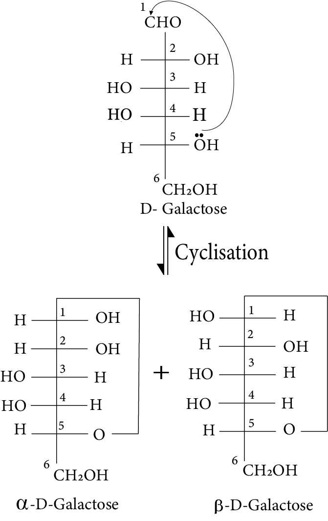
Galactose
Except for C-4, the configurations of all the carbon atoms in \[\beta \]-D-Glucose and \[\beta \]-D-Galactose are the same. Such stereoisomers that differ in configuration at only one carbon other than anomeric carbon are referred to as epimers, and the C atom in question is referred to as an epimeric carbon atom. As a result, \[\beta \]-DGlucose and \[\beta \]-D-Galactose are epimers, and C-4 is an epimeric carbon atom. The linkage in lactose is between C1 of galactose and C4 of glucose. As a result, it also reduces sugar.
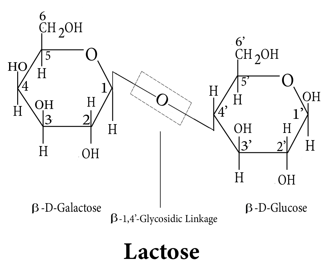
Glycosidic Linkage in Lactose is Between C1 of Galactose and C4 of Glucose
7. Polysaccharides
Polysaccharides are composed of many monosaccharide units linked together by glycosidic linkages. They primarily serve as food storage or structural materials.
7.1 Starch
Plants' primary storage polysaccharide is starch. It is the most important source of nutrition for humans. Cereals, roots, tubers, and some vegetables have a high starch content. It is a -glucose polymer composed of two components: Amylose and Amylopectin. Amylose is a water-soluble component that accounts for about 15-20% of starch. Amylose is a long unbranched chain containing 200-1000 \[\alpha \]-D-(+)-glucose units held together by a C1-C4 glycosidic linkage. Amylopectin is insoluble in water and accounts for approximately 80-85 percent of starch. It is a branched chain polymer of \[\alpha \]-D-glucose units with a chain formed by C1-C4 glycosidic linkage and branching caused by C1-C6 glycosidic linkage.
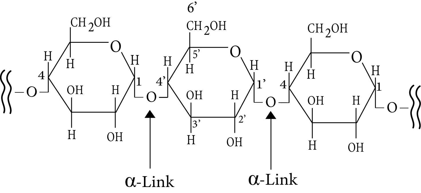
Amylose
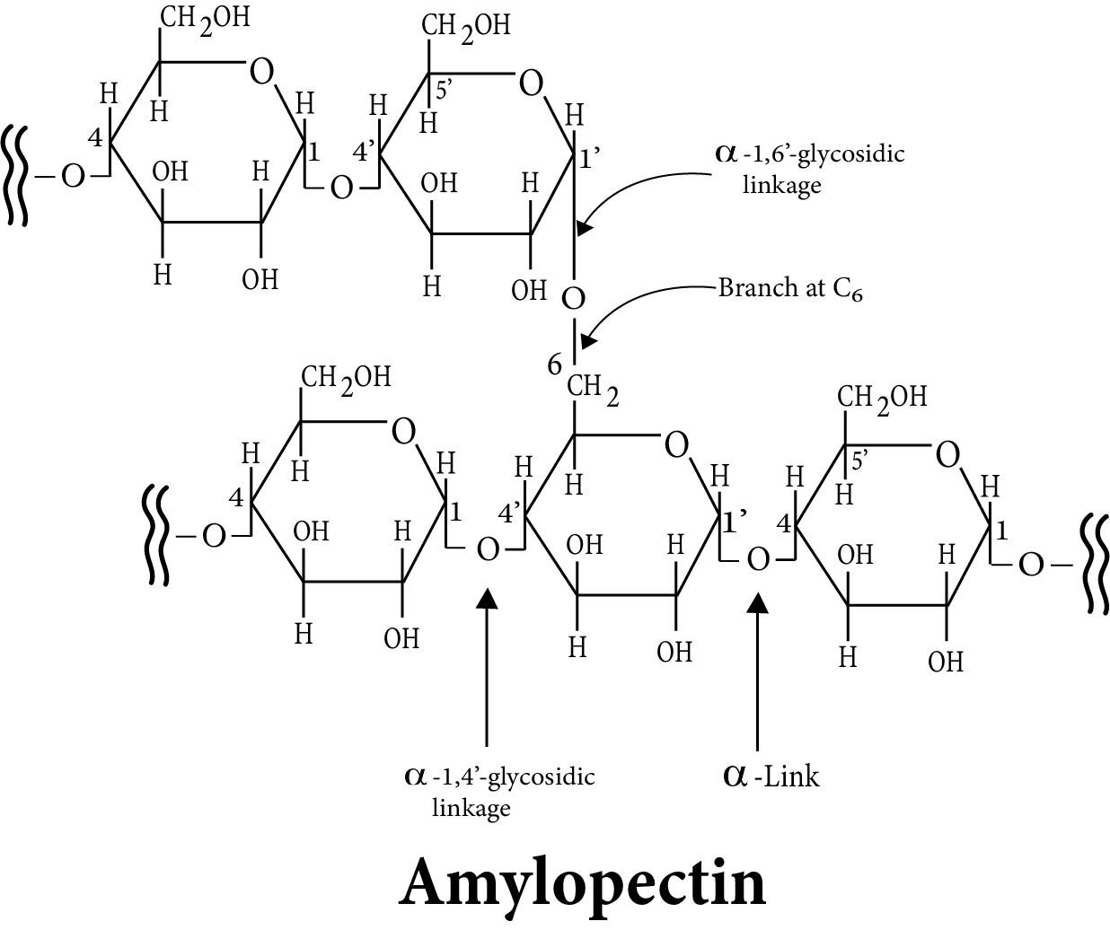
Amylopectin
7.2 Cellulose
Cellulose is found only in plants and is the most abundant organic substance in the plant kingdom. It is a major component of the cell wall of plant cells. Cellulose is a straight-chain polysaccharide made up entirely of \[\beta \]-D glucose units linked together by a glycosidic linkage between C1 of one glucose unit and C4 of the next glucose unit.
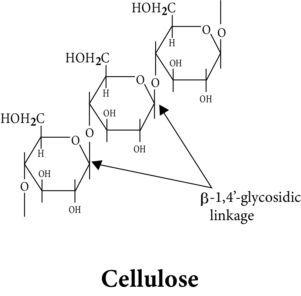
Cellulose
7.3 Glycogen
Carbohydrates are stored as glycogen in the animal body. It is also known as animal starch due to its structure, which is similar to amylopectin but more highly branched. It can be found in the liver, muscles, and brain. When the body requires glucose, enzymes convert glycogen to glucose. Glycogen is found in yeast and fungi as well.
7.4 Summary
Note:
Reducing sugars are all carbohydrates that contain the CHO group, the \[\alpha \]-Hydroxy ketonic group, or the hemiacetal group.
Mutarotation is a phenomenon that occurs in all reducing sugars.
8. Proteins
Protein is derived from the Greek word "proteios," which means "primary" or "of primary importance." Proteins are all polymers of \[\alpha \]-amino acids.
8.1 Amino Acids
Amino acids have both amino (–$NH_2$) and carboxyl (–COOH) functional groups. The amino acids are classified as \[\alpha ,\beta ,\gamma ,\delta \] and so on based on the relative position of the amino group with respect to the carboxyl group. When proteins are hydrolyzed, only \[\alpha \]-amino acids are produced. They may also contain other functional groups.
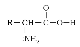
Amino Acids
All \[\alpha \]-amino acids have innocuous names that usually reflect the compound's or its source's property. Glycine is named after its sweet taste (glykos means sweet in Greek), and tyrosine was first obtained from cheese (tyros means cheese in Greek).
8.2 Classification of Amino Acids
The relative number of amino and carboxyl groups in an amino acid molecule determines whether it is acidic, basic, or neutral.
Non-essential amino acids are amino acids that can be synthesized in the body. Essential amino acids, on the other hand, are those that cannot be synthesized in the body and must be obtained through diet.
8.3 Properties of Amino Acids
Amino acids are crystalline, colorless solids. These are water-soluble, high-melting-point solids that act more like salts than simple amines or carboxylic acids. The presence of both acidic (carboxyl group) and basic (amino group) groups in the same molecule causes this behavior.
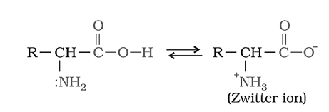
Amino Acids Exhibit Amphoteric Behavior in Zwitter Ionic Form
In aqueous solution, the carboxyl group can lose a proton and the amino group can accept a proton, resulting in zwitter ion, a dipolar ion. This is neutral, but it does have both positive and negative charges. Amino acids exhibit amphoteric behavior in zwitter ionic form, reacting with both acids and bases.
Except for glycine, all other naturally occurring \[\alpha \]-amino acids are optically active due to the asymmetry of the -carbon atom.
These are available in both 'D' and 'L' forms. The majority of naturally occurring amino acids have an L-configuration. The –NH2 group on the left side is used to represent L-amino acids.
8.4.1 Isoelectric Point
An amino acid's isoelectric point (pI) is the pH at which it has no net charge. In other words, it is the pH at which the negative charge on an amino acid exactly balances the positive charge. pI (isoelectric point) = pH at which no net charge exists. The pI of an amino acid with no ionizable side chain, such as alanine, is halfway between its two pKa values. This is due to the fact that at pH = 2.34, half of the molecules have a negatively charged carboxyl group and half have an uncharged carboxyl group, and at pH = 9.69, half of the molecules have a positively charged amino group and half have an uncharged amino group. As the pH rises from 2.34, more molecules' carboxyl groups become negatively charged; as the pH falls from 9.69, more molecules' amino groups become positively charged.
As a result, the number of negatively charged groups equals the number of positively charged groups when the two pKa values are averaged.
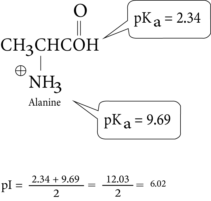
Isoelectric Point of Alanine
If an amino acid has an ionizable side chain, its pI is calculated by averaging the pKa values of similarly ionizing groups (positive ionizing to uncharged or uncharged ionizing to positive). For example, the pI of lysine is the average of the pKa values of two groups that are positively charged in their acidic form but uncharged in their basic form. The pI of glutamate, on the other hand, is the average of the pKa values of the two groups, which are uncharged in their acidic form and negatively charged in their basic form.
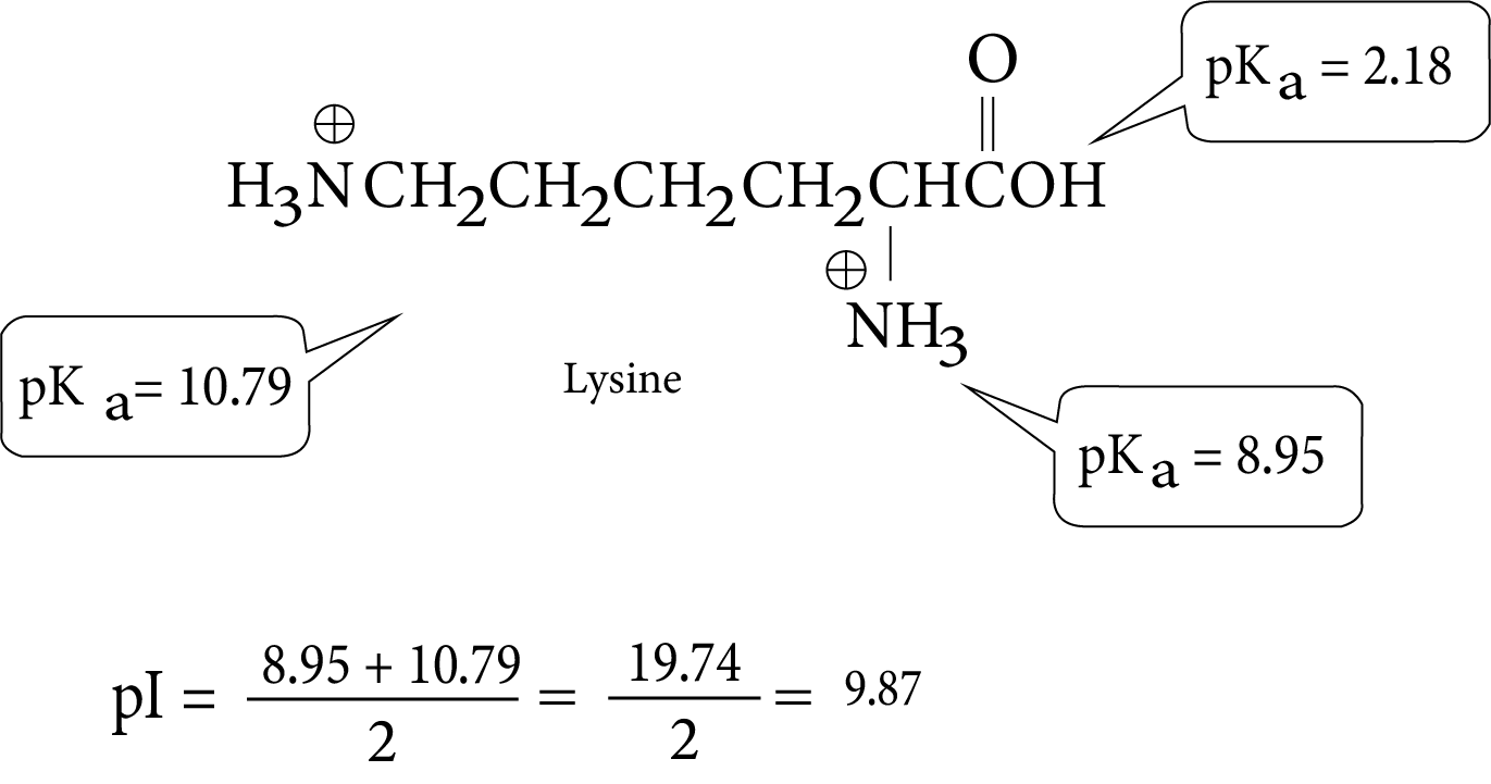
Isoelectric Point of Lysine
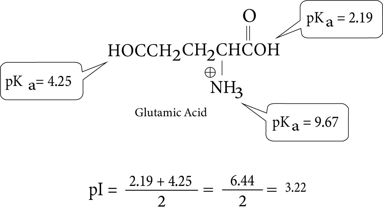
Isoelectric Point of Glutamic Acid
8.5 Structure of Proteins –
The Peptide Bond Proteins are polymers of \[\alpha \]-amino acids that are linked together by peptide bonds or peptide linkage. Peptide linkage is chemically defined as an amide formed between the – COOH group and the –NH2 group.
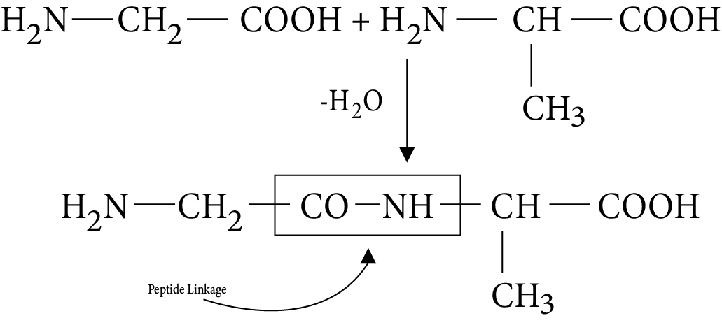
Peptide Linkage in Protein
The reaction between two molecules of similar or different amino acids is initiated by the combination of one molecule's amino group with the carboxyl group of the other. This causes the removal of a water molecule and the formation of a peptide bond –CO–NH–.
8.6 Classification of Proteins
Proteins can be classified into two types on the basis of their molecular shape.
Fibrous Proteins
A fiber like structure is formed when polypeptide chains run parallel and are held together by hydrogen and disulphide bonds. In general, such proteins are insoluble in water.
Keratin (found in hair, wool, and silk) and myosin (found in muscles) are two common examples.
Globular Proteins
This structure is formed when polypeptide chains coil around to form a spherical shape. These are usually water soluble. Insulin and albumin are two common globular proteins. Protein structure and shape can be studied at four different levels: primary, secondary, tertiary, and quaternary.
Primary Structure
One or more polypeptide chains can be found in proteins. Each polypeptide in a protein contains amino acids that are linked together in a specific sequence, and this sequence of amino acids is referred to as the protein's primary structure. Any change in this primary structure, i.e., the amino acid sequence, results in a different protein.
Secondary Structure
A protein's secondary structure is the shape in which a long polypeptide chain can exist. They have been discovered to exist in two different structures: \[\alpha \]-helix and \[\beta \]-pleated sheet. These structures form as a result of the regular folding of the polypeptide chain's backbone caused by hydrogen bonding between the peptide bond's C = O and –NH– groups.
The –NH group of each amino acid residue is hydrogen bonded to the C = O of an adjacent turn of the \[\alpha \]helix in one of the most common ways in which a polypeptide chain forms all possible hydrogen bonds by twisting into a right handed screw (helix).
All peptide chains in \[\beta \]-structure are stretched to nearly maximum extension and then laid side by side, held together by intermolecular hydrogen bonds.
Tertiary Structure
Protein tertiary structure represents overall folding of polypeptide chains, i.e., further folding of the secondary structure. It produces two major molecular shapes: fibrous and globular. Hydrogen bonds, disulphide linkages, van der Waals forces, and electrostatic forces of attraction are the main forces that keep proteins' 2° and 3° structures stable.
Quaternary Structure
Some proteins are made up of two or more polypeptide chains known as sub-units. The relationship of subunits to one another is referred to as quaternary structure.
8.7 Denaturation of Proteins
A native protein is a protein found in a biological system that has a distinct three-dimensional structure and biological activity. When a protein in its native form is subjected to physical change, such as temperature change, or chemical change, such as pH change, the hydrogen bonds are disrupted. As a result, globules unfold, helixes uncoil, and protein loses biological activity. This is referred to as protein denaturation. A common example of denaturation is the coagulation of egg white when heated. Another example is milk curdling, which is caused by the bacteria present in milk producing lactic acid.
9. Nucleic Acids
Every generation of every species resembles its ancestors in a variety of ways. How are these traits passed down from one generation to the next? It has been discovered that the nucleus of a living cell is in charge of the transmission of inherent characteristics, also known as heredity. The particles in the nucleus of the cell that are responsible for heredity are known as chromosomes, and they are made up of proteins and another type of biomolecule known as nucleic acids. These are primarily of two kinds: deoxyribonucleic acid (DNA) and ribonucleic acid (RNA) (RNA). Because nucleic acids are long chain polymers of nucleotides, they are also referred to as polynucleotides.
9.1 Chemical Composition of Nucleic Acids
When DNA (or RNA) is completely hydrolyzed, it produces a pentose sugar, phosphoric acid, and nitrogen-containing heterocyclic compounds (called bases). The sugar moiety in DNA molecules is \[\beta \]-D-2-deoxyribose, whereas the sugar moiety in RNA molecules is \[\beta \]-D-ribose.
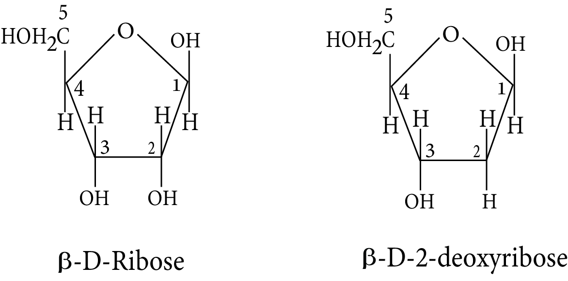
Structure of Beta -D-ribose and Beta -D-2-deoxyribose
Adenine (A), guanine (G), cytosine (C), and thymine are the four bases found in DNA (T). The first three bases of RNA are the same as those of DNA, but the fourth is uracil (U).
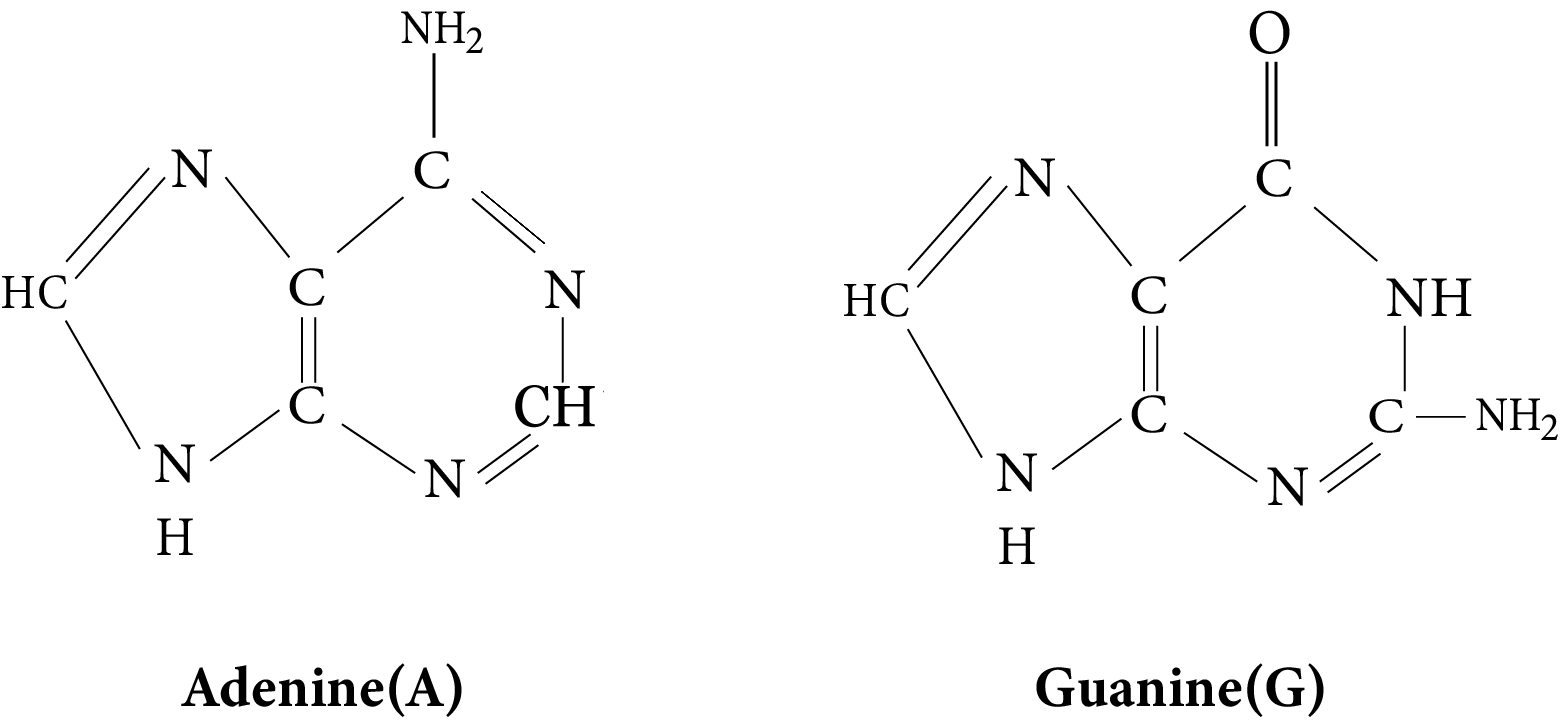
Structure of Adenine (A) and Guanine (G)
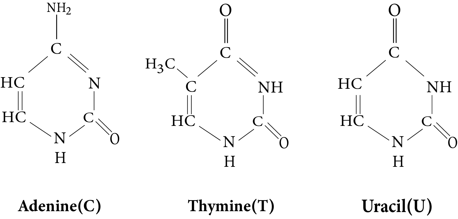
Structure of Cytosine(C), Thymine(T) and Uracil(C)
9.2 Structure of Nucleic Acids
A nucleoside is a unit formed by the attachment of a base to the 1' position of a sugar. The sugar carbons in nucleosides are labeled as 1', 2', 3', and so on. To differentiate these from the bases. A nucleotide is formed when a nucleoside is linked to phosphoric acid at the 5'-position of the sugar moiety.
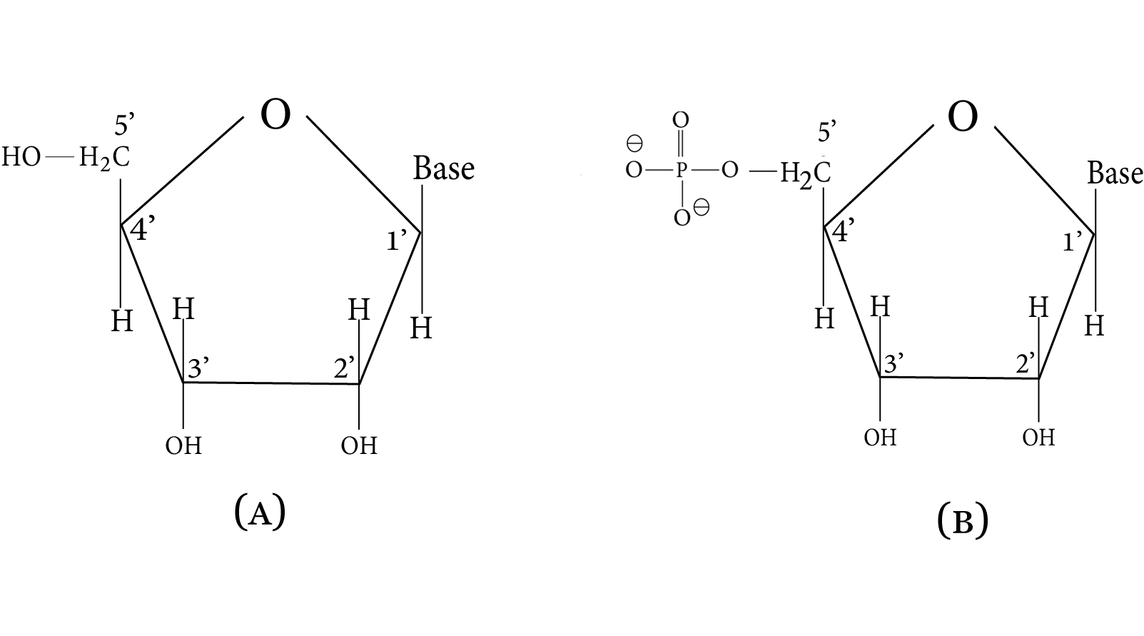
Structure of (a) a Nucleoide and (b) a Nucleotide
The phosphodiester linkage between the pentose sugar's 5' and 3' carbon atoms connects nucleotides. The following is a simplified version of the nucleic acid chain.
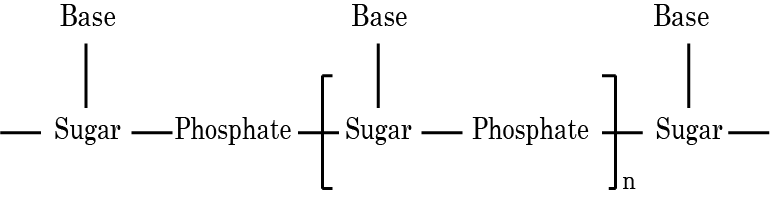
Simplified Version of the Nucleic Acid Chain
The primary structure of a nucleic acid is information about the sequence of nucleotides in its chain. Secondary structure is also present in nucleic acids. The double strand helix structure of DNA was proposed by James Watson and Francis Crick. Two nucleic acid chains are wrapped around each other and held together by hydrogen bonds formed by base pairs. Because hydrogen bonds form between specific pairs of bases, the two strands are complementary to one another. Adenine and thymine form hydrogen bonds, whereas cytosine and guanine form hydrogen bonds. There are helices in the secondary structure of RNA that are only single stranded. They can fold in on themselves to form a double helix structure. There are three types of RNA molecules, each of which serves a different purpose. They are known as messenger RNA (m-RNA), ribosomal RNA (r-RNA), and transfer RNA, respectively (t-RNA).
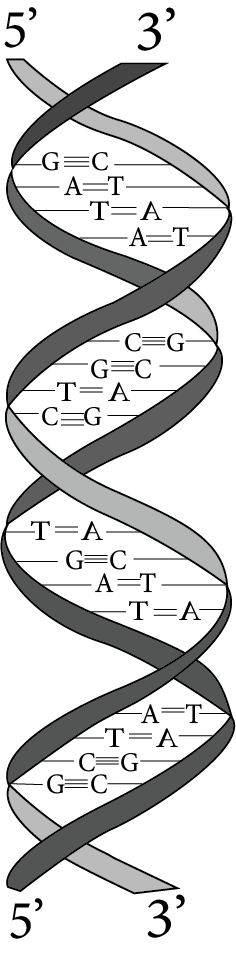
Double Helix Strand of DNA
9.3 DNA Vs RNA
9.4 Biological Functions of Nucleic Acids
DNA is the chemical basis of heredity and can be thought of as a repository of genetic information. Over millions of years, DNA has been solely responsible for maintaining the identity of various species of organisms. During cell division, a DNA molecule can self-replicate, and identical DNA strands are transferred to daughter cells. Protein synthesis in the cell is another important function of nucleic acids. Actually, proteins are synthesised by various RNA molecules in the cell, but the message for a specific protein's synthesis is present in DNA.
10. Enzymes
Enzymes function as biological catalysts. Enzymes are all globular proteins chemically. The following are some important enzymes and their functions.
11. Vitamins
12. Hormones
Hormones are biomolecules that are produced in the ductless (endocrine) glands and transported by the bloodstream to various parts of the body where they control various metabolic processes. These are required in minute amounts and, unlike fats and carbohydrates, are not stored in the body but are constantly produced.
12.1 Steroidal Hormones
12.2 Peptide Hormones
12.3 Amine Hormones
13. Test For Biomolecules
13.1 Test of Carbohydrates
13.1.1 Molish Test
The Molisch test detects all types of carbohydrates, including monosaccharides, disaccharides, and polysaccharides. Molisch reagent (1 percent alcoholic solution of \[\alpha \]-naphthol) is added to an aqueous carbohydrate solution, followed by conc. \[{{\text{H}}_{\text{2}}}{\text{S}}{{\text{O}}_{\text{4}}}\] along the test tube's sides. At the intersection of the two layers, a violet ring forms.
13.2 Test for Proteins
13.2.1 Biuret Test
An alkaline solution of a protein when treated with a few drops of 1% \[{\text{CuS}}{{\text{O}}_{\text{4}}}\] solution, produces a violet colouration. The colour is due to the formation of a coordination complex of \[{\text{C}}{{\text{u}}^{{\text{ + 2}}}}\] with carbonyl group and –NH– groups of the peptide linkages.
13.2.2 Xanthoproteic Test
When a protein is treated with concentrated \[{\text{HN}}{{\text{O}}_{\text{3}}}{\text{,}}\] it turns yellow. This test is performed by a protein composed of \[\alpha \]-amino acids containing a benzene ring, such as tyrosine, phenylalanine, and others, and the yellow color is caused by benzene ring nitration. An important example of this test is when concentrated \[{\text{HN}}{{\text{O}}_{\text{3}}}{\text{,}}\] is spilled on your hands, the skin turns yellow due to nitration of the benzene ring of the amino acids in your skin's proteins.
13.2.3 Millon’s Test
Millon's reagent is a nitric acid solution of mercurous nitrate and mercuric nitrate with little nitrous acid. When Millon's reagent is added to an aqueous protein solution, a white ppt. is formed. All proteins containing phenolic \[\alpha \]-amino acids, such as tyrosine, pass this test. As a result, gelatin that does not contain phenolic \[\alpha \]-amino acids fails this test.
13.2.4 Ninhydrin Test
A blue-violet color is produced when proteins are boiled in a dilute aqueous solution of ninhydrin (2, 2-dihydroxyindane-1,3-dione). This test is provided by all \[\alpha \]-amino acids. Proteins and peptides both pass this test because they give \[\alpha \]-amino acids when hydrolyzed.
Class 12 Chemistry Chapter 10 Important Topics and Subtopics Covered
Class 12 Chemistry Chapters 10 Details, Formulas and Concepts.
Classification of Biomolecules: Biomolecules are classified into four main categories based on their chemical nature and functions:
Carbohydrates: Sugars, starches, cellulose, etc.
Proteins: Polymers of amino acids.
Lipids: Fats, oils, phospholipids, etc.
Nucleic Acids: DNA, RNA, ATP, etc.
Primary Structure of Proteins: The primary structure of a protein refers to the sequence of amino acids in the polypeptide chain. It is determined by the order of amino acids linked by peptide bonds.
Carbohydrate Chemistry: Key concepts in carbohydrate chemistry include:
Monosaccharides: Simple sugars such as glucose, fructose, and galactose.
Disaccharides: Two monosaccharides linked by a glycosidic bond, such as sucrose, lactose, and maltose.
Polysaccharides: Complex carbohydrates formed by the polymerization of monosaccharide units, such as starch, glycogen, and cellulose.
Enzyme Kinetics: Enzymes are biological catalysts that increase the rate of biochemical reactions.
Nucleic Acid Structure: Nucleic acids are polymers of nucleotides and include DNA and RNA. Key concepts include:
DNA Structure: Double helix structure composed of two complementary strands of nucleotides held together by hydrogen bonds.
RNA Structure: Single-stranded molecule involved in protein synthesis and gene expression.
Importance of Revision Notes for Class 12 Chemistry Chapter 10
Summarises Key Points: Condenses important concepts for quick review.
Saves Time: Provides a fast way to revise before exams.
Highlights Essentials: Focuses on crucial topics and definitions.
Improves Memory: Helps in better retention of information.
Enhances Exam Prep: Targets weak areas for more effective study.
Clarifies Concepts: Simplifies complex ideas for easier understanding.
Includes Visuals: Uses diagrams and charts for better grasp.
Boosts Confidence: Prepares students thoroughly for exams.
Tips For Learning the Class 12 Chapter 10
Focus on core processes with illustrations and examples.
Draw and label diagrams for clarity.
Create summaries of each process.
Connect concepts to everyday examples.
Solve past exam questions to test understanding.
Explain concepts to others to reinforce learning.
Revisit material frequently to retain information.
Conclusion
The chapter on Biomolecules in Class 12 Chemistry notes provides a comprehensive overview of the essential organic compounds that drive biological processes. By studying carbohydrates, proteins, lipids, and nucleic acids, students gain insights into their structures, functions, and importance. This foundational knowledge is critical for understanding more complex biological systems and processes, making it an integral part of the chemistry curriculum.
Related Study Materials for Class 12 Chapter 10
Revision Notes Links for Class 12 Chemistry Revision Notes
Related Study Material Links for Class 12 Chemistry
FAQs on Biomolecules Class 12 Chemistry Chapter 10 CBSE Notes - 2025-26
1. What is the most effective sequence for revising Biomolecules in Class 12 Chemistry using summary notes?
Start your revision by understanding the classification of biomolecules into carbohydrates, proteins, lipids, and nucleic acids. Next, review the structure and types of carbohydrates (monosaccharides, disaccharides, polysaccharides), focusing on their functions and reactions. Then, move on to proteins, covering amino acid structure, peptide bonds, and the four levels of protein structure. Follow with lipids, looking at their types, structure, and saponification reactions. Finally, study nucleic acids, emphasizing DNA and RNA structures, replication, and biological roles. This sequence ensures logical flow and thorough coverage as per the CBSE 2025–26 syllabus.
2. How do concise revision notes improve retention and exam performance for Chapter 10?
Concise revision notes enhance retention by summarizing core concepts, key definitions, and essential formulas in an easy-to-review format. These notes save time, minimize information overload, and help students focus on high-weightage topics. By highlighting vital details and using diagrams or concept maps, students can quickly revisit important points before exams, leading to improved memory, understanding, and confidence while answering application-based questions.
3. Which core ideas and definitions should be prioritized when revising Biomolecules for Class 12 Chemistry?
Prioritize the following core ideas:
- Classification of biomolecules (carbohydrates, proteins, lipids, nucleic acids)
- Key definitions: monosaccharides, disaccharides, polysaccharides, peptide bond, nucleotide, mutarotation
- Structures and functions of glucose, starch, cellulose, DNA, and RNA
- The four levels of protein structure (primary, secondary, tertiary, quaternary)
- Common reactions—hydrolysis, saponification, peptide bond formation
- Role and significance of biomolecules in living systems
4. How can students use concept maps to connect topics while revising Biomolecules?
Creating concept maps helps visually link related topics—for example, mapping the relationship between types of carbohydrates, their structure, and biological function. Linking protein structure levels to their roles, or connecting DNA structure with genetic information flow, makes interconnections clear. These visual tools aid memory retention, clarify how ideas are related, and allow for efficient last-minute revision.
5. What are common misconceptions students face when studying biomolecules and how can these be avoided through notes?
Students often believe all sugars are reducing, confuse DNA with RNA functions, or overlook the difference between peptide and glycosidic bonds. They might also think all proteins have similar structures. Summarized notes should clearly distinguish reducing vs non-reducing sugars, specify the roles of DNA vs RNA, and explain different bond types and protein structure levels to address these misconceptions.
6. Why is understanding the four levels of protein structure crucial during rapid revision?
Recognizing primary, secondary, tertiary, and quaternary protein structures is essential because each level determines the protein's function, stability, and ability to interact in biological processes. For example, denaturation affects secondary and tertiary structures, impacting enzyme activity. Quick recall of these distinctions supports strong answers to application-based and reasoning questions in board exams.
7. How should real-life examples be used in summarising the functions of different biomolecules?
Link each biomolecule to a specific function and real-life context: starch is an energy reserve in plants, cellulose gives structural support to plant cell walls, glycogen stores energy in animals, and insulin regulates blood sugar. Using relatable examples makes it easier to remember both structure and function during revision.
8. How do summary notes address application-based and HOTS questions in Biomolecules?
Well-organized summary notes not only cover factual content but also include conceptual connections, flowcharts, or diagrams that clarify structure–function relationships and biological significance. By including example-based questions or 'reasoning why' a process occurs, notes prepare students for higher-order thinking and application-based CBSE 2025–26 exam items.
9. What revision strategies are recommended for mastering the detailed reactions and structures within Biomolecules?
Use repetition and stepwise writing to memorize reactions (such as hydrolysis of sucrose, peptide bond formation, saponification). Draw labelled diagrams for the structures of glucose, amino acids, or DNA. Break complex reactions into simple, logical steps and revise them regularly for long-term retention. Practice explaining these reactions out loud or teaching them to someone else for deeper understanding.
10. How can students ensure their revision covers all essential Biomolecules concepts as outlined in the CBSE/NCERT 2025–26 syllabus?
Cross-check their summary notes with the latest official CBSE/NCERT Chemistry syllabus. Make sure each biomolecule type (carbohydrates, proteins, lipids, nucleic acids) is included, along with their definitions, structures, main reactions, and biological roles. Prioritize concepts mentioned in the chapter overview and highlighted in the official syllabus to ensure nothing is missed before the exam.