

How to Prepare and Observe a Temporary Leaf Peel Mount for Exam Success 2025-26
Biology experiment - Experiment preparing a Temporary Mount of a Leaf Peel to Show Stomata
Plants are considered as living organisms because they can perform the functions of growth, development and reproduction. One such quality that the plants have which is similar to animals is the ability to perform gaseous exchange. This gaseous exchange in plants is done by tiny pores known as stomata. Stomata are present on the leaves where photosynthesis takes place. This is an advanced feature which is found in higher plants such as gymnosperms and angiosperms.
Table of Contents
The following article contains: -
Aim
Requirements
Theory
Methodology
Observations
Conclusion
Aim
To prepare a temporary mount of a leaf peel to show the presence of stomata.
Requirements
Leaf, forceps, blade, glycerin, slides, coverslips, compound microscope, water, safranin, blotting paper, etc.
Theory
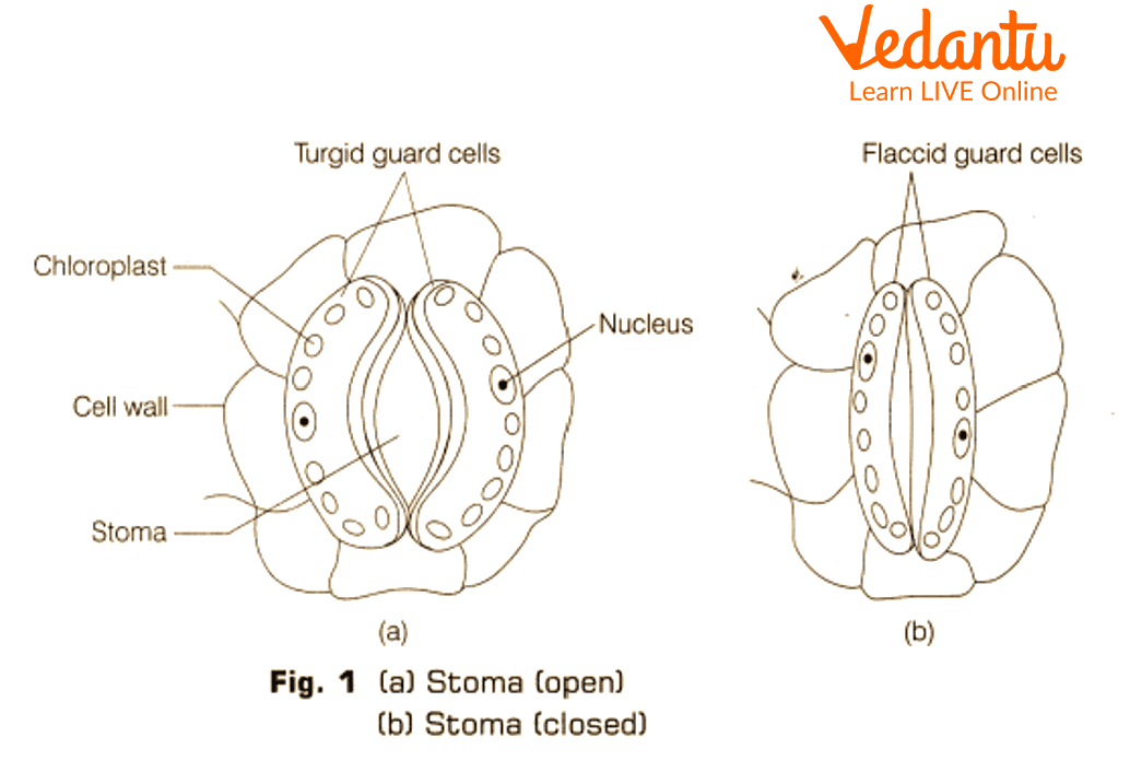
Stomata are small pores which are guarded by kidney shaped or dumb bell-shaped guard cells. These guard cells absorb water and swell as a result the stomata opens up; when water is lost from the guard cells they turn flaccid and the pores close. These guard cells contain chloroplast and nucleus. Stomata is present in large numbers on the lower epidermis in dicot leaf and are uniformly distributed between both surfaces in monocots.
Procedure
For this leaf activity, take fresh leaves from any plant and clean and dry it.
Flip the leaf on the lower side and tear the leaf tangentially i.e., at an angle. Due to this process a transparent tissue is seen which contains the stomata.
Take the leaf peel with forceps in a petri dish and add a few drops of safranin to it.
Keep the peel in the stain for 2-5 mins so the stain can be absorbed by the peel.
Place this stained leaf peel on a glass slide.
We generally mount the material in the slide after adding a drop of glycerin over it and then gently placing a coverslip.
Clean excess glycerin using a blotting paper.
Place the slide under a compound microscope and visualize the leaf peel first at low power and then at high power.
Record the observations.
Observations
At low power of the microscope epidermal cells were visible which were a single layer and do not contain any intercellular spaces.
Scattered throughout the epidermal cells were small tiny pore-like structures.
At high power these tiny pores appear with proper stomatal openings.
These pores were surrounded by two kidney-shaped guard cells which control the opening and closing of the stomatal pore.
Each guard cell contained one nucleus with multiple chloroplasts.
Result
In this leaf activity the stomata were observed on the lower epidermis of the leaf. These pores contain kidney shaped guard cells, hence it can be concluded that this leaf was a dicot leaf. The guard cells contain a nucleus and chloroplasts.

This diagram shows the microscopic view of a dicot stomata.
Precautions
Do not over stain the specimen.
Use an adequate amount of glycerin.
Clean the slide properly before putting it under the microscope.
Lab Manual Questions
1. Why is the temporary mount slide mounted in glycerine?
Ans: In general mounting of material in the slide is done using glycerin. Glycerin has the capacity to remain moist for a long time and hence prevents the dehydration of the sample.
2. How to differentiate between guard cells and epidermal cells?
Ans: Epidermal cells are irregular in shape, have no intercellular spaces and lack chloroplast. Guard cells are specialized cells and they are either kidney or dumb belled shaped, They are green as they contain many chloroplasts.
3. How is the distribution of stomata in monocot leaves?
Ans: In monocot leaves, the stomata is equally distributed on lower and upper epidermis as they contain equal amounts of chloroplast and both surfaces perform equal amounts of photosynthesis.
4. What is the shape and function of guard cells?
Ans: In dicot leaves the guard cells are kidney shaped, whereas in monocot leaves the guard cells are dumb-belled shaped. The function of guard cells is to protect the tiny pores and control the opening and closure of the stomata.
Viva Questions
Why is safranin used for staining plant cells?
Ans: Safranin is a red colored stain and stains lignin, suberin and other plant parts very easily.
What is the shape of guard cells when seen under a microscope?
Ans: In monocot plant guard cells are dumb belled shaped; in dicot plant guard cells are kidney shaped.
Which instrument can be used to know the size of stomata?
Ans: A porometer is used to know the size of the stomata.
Which plant does not contain stomata?
Ans: Hydrilla does not contain stomata.
What is the modification of stomata in gymnosperms and xerophytic plants?
Ans: In gymnosperms and xerophytic plants the stomata are sunken.
What are the functions of a leaf?
Ans: Leaf performs the functions of Photosynthesis, Respiration and Transpiration.
Why is transpiration required by plants?
Ans: Transpiration helps in building a transpiration pull in plants, this helps in the movement of water from roots to the top leaves ( movement against gravity).
Give examples of plants having kidney shaped stomata.
Ans: Banyan, Betel leaf plant, Peepal, Mango, False Ashoka etc.
Give examples of plants having dumb- belled shaped stomata.
Ans: Banana, Grasses, Rice, Jowar, Bajra, etc.
Which part of the stomata contains chloroplast?
Ans: The guard cells of the stomata contain chloroplast.
Practical Based Questions
For the above leaf activity, we generally mount the material in the slide using _____
Water
Glycerin
Alcohol
Wax
Ans: Glycerin
Find the odd man out
Guard cells
Epidermal cells
Stomata
Trichomes
Ans: Trichomes
Which of the following is not a function of stomata?
Transpiration
Respiration
Photosynthesis
Gaseous exchange
Ans: Photosynthesis
Hormone responsible for closing of stomatal pore
Abscisic acid
Auxins
Gibberellins
Cytokinin
Ans: Abscisic acid
Inorganic ion assisting in opening and closing of the stomata are
Ca+ ions
Na+ ions
K+ ions
Cl- ions
Ans: K+ ions
A temporary mount of a tissue is made in which of the following has to be used?
Safranin and glycerin
Acetocarmine and alcohol
Iodine and water
Safranin and water
Ans: Safranin and glycerin
Hormone responsible for opening of stomatal pore
Abscisic acid
Cytokinin
Gibberellins
Auxins
Ans: Cytokinin
In an experiment a temporary mount of a leaf peel is prepared from betel leaf to show stomata, from where the peel should be taken?
Petiole of the leaf
Vein of the leaf
Lower surface of the leaf
Upper surface of leaf
Ans: Lower surface of the leaf
Regarding the distribution of stomata in monocot leaves which of the following is true?
In monocots, leaves are amphistomatic
Monocot leaves stomata are situated only on the dorsal surface
Grasses have stomata only on upper surface
Monocot stomata are large and kidney shaped
Ans: In monocots, leaves are amphistomatic
Which part of the leaf peel is pink colored after staining with safranin?
Stomata only
Epidermal cells
All parts of the peel
Only the nucleus
Ans: All parts of the peel
Which of the following is used to see the stomata
Electron microscope
Compound microscope
X-ray microscope
Simple microscope
Ans: Compound microscope.
Summary
Stomata is the most important part of the plant, as it helps in exchange of gases and facilitates most vital processes of the plants i.e., photosynthesis, respiration and transpiration. A stoma contains a stomatal pore, guard cells, subsidiary cells and epidermal cells.
FAQs on Step-by-Step Guide to Stomata Observation Using Leaf Peels: Class 10 Biology Practical Demo
1. Lower epidermis contains more stomata in dicot leaves. Explain.
Dicots plants are tall trees which are exposed to excessive sunlight. Lots of water will be lost from the stomatal pores due to transpiration if stomata are present on the upper epidermis. Hence to prevent, excessive transpiration and dehydration of plant stomata is present more over the lower epidermis.
2. Are stomata present on roots? If not then explain why.
Stomata are present where gaseous exchange takes place and hence is seen only on the aerial parts of the plant such as leaves. They are not present on roots as roots are buried in the soil and there no gaseous exchange takes place.
3. What is the mechanism of opening and closing of stomatal pore?
Stomatal pore open and closing is controlled by the guard cells. These cells contain differential thickenings over the inside and outside walls. The inner wall is thick, less elastic and the outer walls are thin, highly elastic. Hence when water reaches these cells, they become turgid and open the pore, and when water is lost, they become flaccid and pores close.
4. Differentiate between a monocot plant and dicot plant based on the leaf?
Based on leaf structure a monocot and dicot can be differentiated-
Monocot leaf | Dicot leaf |
Leaf shows parallel venation. | Leaf shows reticulate venation. |
Dumb belled shaped guard cells. | Kidney shaped guard cells. |
Stomata is equally distributed on both epidermis. | Stomata majorly present on the lower epidermis. |


