


Key Insights into Blastula Structure for 2025-26 Exam Success
You might be knowing that after 7-9 days of fertilisation, zygote implants in the uterus. The zygote undergoes repeated divisions called cleavage. Cleavage includes a series of successive mitotic divisions that transform the zygote into an embryo. In this experiment, we will observe many characteristics of the human blastula stage. Blastula formed after 5-6 days of fertilisation. And it implants in a uterine wall process known as implantation. In this article, we will also describe the structure of the blastula.
Table of Content
Aim
Articles Required
Theory
Procedure
Observations
Results
Precautions
Lab Manual Questions
Viva Questions
Practical Based Questions
Conclusion
Aim
This experiment aims to study the T.S of Blastula through permanent slides.
Articles Required
Permanent slides
Compound microscope
Theory
The blastula is an important stage of embryonic development. Zygote forms after male and female gamete fertilisation. Then zygotes undergo further cleavages to form embryos. Cleavage occurs in the upper part of the fallopian tube. After some cleavages, a solid mass of about 8-16 cells formed; this mass is called a morula. Then the morula undergoes further cleavages to form a blastula. The blastula contains about 64 cells. There are six types of blastula.
The outer layer of the blastula is called the blastoderm, and the inner cavity is called the blastula, undergoes further division or development and forms a gastrula. The process of formation of a gastrula from blastula is known as gastrulation. The T.S. of the gastrula of the frog will be discussed in further experiments
Procedure
Fix the slides under the microscope properly. Then observe the slide under a low-power microscope and then observe it under a high-power microscope.
Observations
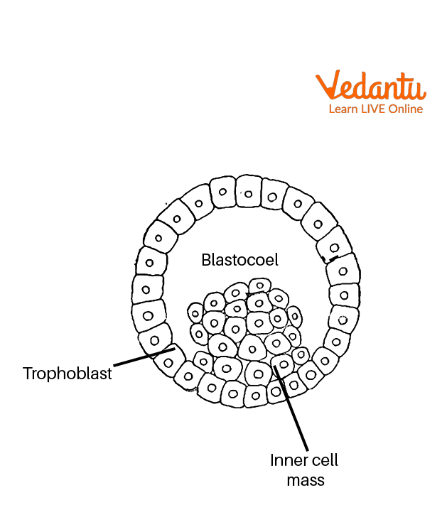
In a microscope, we can see a large spherical mass of 64 cells. This mass is the blastula stage of the embryo of mammals.
The outer layer of this mass is trophoblast or trophectoderm and inside this inner cell, mass is present. After these inner cell masses convert into three different germ layers which will make a complete organism.
Blastocoel is the fluid-filled cavity present within the envelope. The embryonic pole is the pole of the trophoblast through which inner cell mass is attached, while the embryonic is the opposite pole.
The inner cell mass is the precursor of the embryo.
Blastula Diagram
Results
Different parts of the blastula such as the trophoblast and inner cell mass are seen in T.S of the blastula slide.
Precautions
Always first focus slides under a low power microscope and then after under a high power microscope.
And use fine adjustment while focusing slides under the microscope.
Always clean slides before doing an experiment.
Handle slides very carefully.
Lab Manual Questions
Q1. What is morula?
Ans: It is a stage of embryogenesis in which a solid mass is formed of about 16 cells.
Q2. What is implantation?
Ans: Implantation is the attachment of the blastocyst to the uterine wall.
Q3. When does implantation occur in human beings?
Ans: Implantation occurs in human beings after 7 days of fertilisation.
Viva Questions
Q1. Is morula a 16-celled stage?
Ans: Yes, morula is a 16-celled stage. Zygote undergoes cleavage to form a morula. It is mulberry shaped and divides further to form blastula.
Q2. What is cleavage in Biology?
Ans: Cleavage is a kind of mitotic division in which a cell is divided into a large number of cells and overall volume remains the same but per cell volume decreases.
Q3. What is the difference between division and cleavage?
Ans: The main difference between division and cleavage is that in cleavage size/volume of cell is reduced after division but in division size of progeny cell remains same as parent cell.
Q4. What is blastula used for?
Ans: Blastula formed by division in zygote, it later develops into blastocyst which further undergoes development to form embryos.
Q5. What are the types of blastula?
Ans: There are many types of the blastula. Some types are- coeloblastula, periblastula, discoblastula, amphiblastula, and stereoblastula.
Q6. What is LH surge?
Ans: Sudden increase level of LH hormone is known as an LH surge. It is important for ovulation. Ovum is released after 36-40 hours of blood level of luteinising hormone.
Q7. Which hormone is released from Graafian follicles?
Ans: Graafian follicles mature into Graafian follicles. During development, it releases oestrogen hormone, which helps in the further development of follicles.
Q8. What is embryogenesis?
Ans: Embryogenesis is the process of the formation of an embryo from zygote. It is a process by which an embryo matures into a foetus. This process occurs after fertilisation.
Q9. What are the different stages of embryogenesis?
Ans: The different stages of embryogenesis are- fertilisation, blastocyst formation, blastocyst implantation, embryo development, and foetus development.
Practical Based Questions
Q1. What cell stage is blastula?
First stage
Second stage
Third stage
Fourth stage
Ans: 2. Second stage
Q2. How many-celled stages is a blastula?
32-64 celled stage
128 celled stage
12-16 celled stage
None of the above
Ans: 1. 32-64 celled stage
Q3. Which is the part of the blastula?
Trophoblast
Ectoderm
All of the above
None of the above
Ans: 1. Trophoblast
Q4. Which cavity is present in the blastula?
Gastrocoel
Gastrula
Blastocoel
None of the above
Ans: 3. Blastocoel
Q5. What is the fate of blastula?
Formation of gastrula
Formation of morula
Formation of zygote
None of the above
Ans: 1. Formation of gastrula
Q6. How many cells are in the morula stage?
8-16 cells
12-14 cells
32-64 cells
None of the above
Ans: 1 . 8-16 cells.
Q7. Is blastula bigger than zygote?
Yes
No
Of the same size
None of the above
Ans: 3. Of the same size
Q8. Which kingdom has a blastula stage?
Plantae
Mineral
Animalia
None of the above
Ans: 3. Animalia
Conclusion
In this article, we have studied an experiment on the ts of blastula under a microscope. The blastula is a 64-celled stage divided into two parts. The inner part is known as the inner cell mass consists of a large number of cells, whereas the outer layer is known as the trophoblast. Blastocysts undergo implantation in the uterus. There are various types of blastula depending on many factors. Blastula undergoes further development to form gastrula process known as gastrulation.
FAQs on Master the Transverse Section of Blastula: Class 12 Biology Guide for 2025-26
1. What are the most important structural features to identify in the T.S of blastula through permanent slides for CBSE 2025-26 Biology exams?
When examining the transverse section (T.S) of a blastula, focus on identifying the following key structures as per CBSE syllabus:
- Trophoblast (trophectoderm) – outer cell layer
- Inner cell mass – cluster of cells inside
- Blastocoel – fluid-filled cavity
- Cell arrangement (about 64 cells)
2. How is the process of cleavage significant in the formation of a blastula, and why is this asked frequently in exams?
Cleavage is a rapid series of mitotic divisions following fertilization, transforming the zygote into a multicellular structure without increasing overall volume. This process is fundamental in forming the morula (16-cell stage) and then the blastula. It is frequently asked as it covers both conceptual understanding and application, making it important for both short and long exam answers.
3. Explain the steps to properly focus a permanent slide of blastula under a compound microscope for board practicals.
To focus a blastula permanent slide:
- Clean the slide before use.
- Place the slide firmly on the stage and secure with clips.
- Start with low power objective and bring into focus using coarse adjustment.
- Switch to high power objective for detailed observation, using fine adjustment.
- Handle slides carefully to prevent breakage.
4. What types of high-order thinking (HOTS) questions can be expected about differences between blastula and morula in CBSE board exams?
Board papers may require you to differentiate between blastula and morula. Key areas include:
- Morula: 8–16 cells, solid ball, no cavity
- Blastula: 64 cells (typically), presence of blastocoel (cavity), differentiated into cell layers
- Developmental progression: Morula precedes blastula
5. Why is the blastula stage critical for further embryonic development, and how can this be framed as a 5-mark exam question?
The blastula stage is essential because it establishes the basic cellular organization and spatial arrangement needed for gastrulation. During gastrulation, the three germ layers form (ectoderm, mesoderm, endoderm), which give rise to all tissues and organs. Exam questions may require a detailed explanation of the significance of blastula in development, focusing on cell differentiation and future tissue specialization for 5-mark answers.
6. Discuss the various types of blastula and their importance, as may be asked in long-form CBSE questions.
Types of blastula are based on the structure and presence of cavities, including:
- Coeloblastula: Hollow with a blastocoel; seen in echinoderms
- Stereoblastula: Solid, no cavity; found in certain invertebrates
- Periblastula: Central yolk, peripheral nuclei; typical in insects
- Discoblastula: Disc of cells atop yolk; seen in reptiles and birds
7. What are some common practical mistakes to avoid during the observation of the blastula slide, relevant for internal assessments?
Common errors include:
- Not cleaning the slide before use
- Using high power directly without initial low power focusing
- Rough handling causing slide breakage
- Poor adjustment resulting in unclear images
8. How can a diagram-based question on T.S of blastula be answered effectively for maximum marks in CBSE board exams?
For diagram-based questions:
- Neatly draw the T.S. showing trophoblast, inner cell mass, and blastocoel
- Label each part clearly
- Provide a concise caption
- Mention the significance of each part, if asked
9. What is the fate of the blastula, and how might application-based questions use this concept in board exams?
The blastula undergoes further development to become a gastrula through gastrulation. Application questions can link the fate of the blastula to real-life scenarios such as developmental disorders, or ask you to predict outcomes if gastrulation fails, testing deep understanding for higher-order marks.
10. Why is it important for students to understand the marking weightage and frequent board trends related to the T.S of blastula practical and theory questions?
Understanding marking weightage helps prioritize study, as T.S. of blastula often appears as a short answer in theory (2–3 marks), or a diagram/identification in the practical (3–5 marks). Recognizing board trends ensures efficient preparation, helping students focus on scoring areas and frequently asked exam questions for CBSE Biology 2025–26.